Accuracy of Mandibular Rami Measurements in Prediction of Sex
- *Corresponding Author:
- Abhishek Singh Nayyar
Saraswati-Dhanwantari Dental College and Hospital and Post-Graduate Research Institute, Parbhani, Maharashtra, India
Tel: 073509 90780
E-mail: singhabhishek.rims@gmail.com
This is an open access article distributed under the terms of the Creative Commons Attribution-Non Commercial-ShareAlike 3.0 License, which allows others to remix, tweak, and build upon the work non-commercially, as long as the author is credited and the new creations are licensed under the identical terms.
Abstract
Background: Sex of an individual can be determined by means of skeletal indicators when soft tissues are not available for analysis. Also, when entire skull is not available for analysis, mandible plays a vital role in prediction of sex. As various studies have proven the accuracy of panoramic radiographs in providing anatomical measurements, the present study was conducted using digital orthopantomographs (OPGs) in the South Indian population for the same. Aim: To measure and compare various measurements of the ramus in mandible on digital orthopantomographs (OPGs) and to assess the usefulness of such measurements in prediction of sex in an individual. Materials and Methods: A cross-sectional, observational study was carriedout using 500 digital orthopantomographs (OPGs) with five rami measurements taken for each radiograph in the South Indian population. The determination of sex was done by discriminant function analysis with a prediction accuracy being 84.1% amongst females and 76% amongst males. Results: All the variables were found to be good predictors for prediction of sex in the study with Condylar height/Maximum ramus height; Projective height of ramus; and Coronoid height, being highly significant. Conclusion: This study on rami measurements showed that significant sex-related dimorphism is evident in rami of mandibles indicating its potential usage in mass disasters and otherwise in prediction of sex in individuals with disputed identity.
Keywords
Mandibular ramus, Digital Orthopantomographs (OPGs), Prediction of sex
Introduction
In world, where terrorism and crime rule the roost, the investigative measures should be potent enough to identify the victims. Even the minutest remains from the body, helpful for identification, should not be neglected because even the oral mucosal cells collected from tooth brushes have been used to identify victims of world trade centre attack. In such a context, forensic medicine investigative methods need to be highlighted [1].
Also, total number of affected and killed population is significantly increasing day by day in mass disasters. The World Disaster Report published by International Federation Of Red Cross And Red Cross Societies in 2012 showed that the total number of people affected and killed in mass disasters in 2011 itself was around 17 crores and 30 thousands respectively [2]. This considerably higher number of affected population signifies the role of forensic anthropologists in such mass disasters.
The role of an anthropologist is to create a biological profile of unknown skeletal remains, to arrive at conclusions regarding its age, sex, stature and so on that might lead to personal identification. Prediction of sex is one of the leading questions when formulating the biological profile of an individual [3].
There are several methods for prediction of sex. Rosing et al. recommended a first evaluation phase by morphologic character before moving to the second phase of molecular analysis [4,5].
When soft parts are unavailable, prediction of sex can be based only on the characters of the skeleton [1,6].
Skull is one of the easily sexed portions of the human skeleton. In cases of mass disasters, the entire skull is not readily available for analysis and the technical procedure has to be based on the fragmented bones of the skull. In such cases, mandible plays a vital role in prediction of sex in an individual. Humphery et al. stated that sexual dimorphism is reflected in mandibular ramus than the body [7,8].
Radiographic examination plays a significant role in diagnosing non-accidental injuries in children, in medical negligence and in establishing biological aging in the disputed cases.9 Various workers have claimed that prediction of sex by radiographic study of skulls is a reliable method which provides an accuracy of up to 80-100%.1,10,11 Hence, this study aimed at evaluating the reliability of mandibular rami in prediction of sex in the South Indian population and purpose the use of same in forensic analysis and anthropology.
Materials and Methods
This was a cross-sectional, observational study conducted in the Department of Oral Medicine and Radiology using 500 digital orthopantomographs (OPGs) taken with the help of SIRONA digital panoramic and cephalometric system in the South Indian population in the age range of 20 to 60 years who visited the Department for their routine radiographic examination after approval from the Institutional Ethical Committee.
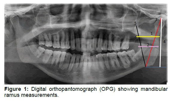
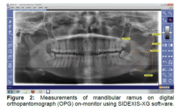
Pathological, fractured and deformed mandibles were excluded from the study. Mandibular rami measurements were carried-out using SIDEXIS-XG software. Radiographs of the patients who were not having pathological, fractured or, deformed mandibles were collected and stored along with their demographic data. Each image was imported into the SIDEXIS-XG software. The tool “Measure Length” was selected in the software and two points were selected using the mouse driven method between which the length was displayed by the software (Figures 1 and 2).
Variable A: Maximum ramus width: Distance between most anterior and posterior points on the mandibular ramus depicted as 1 in Figure 1;
Variable B: Minimum ramus width: Smallest antero-posterior diameter of the mandibular ramus depicted as 2 in Figure 1;.
Variable C: Condylar height / Maximum ramus height: Distance between the most superior points on the condyle to the most protruded point on inferior border of the ramus depicted as 3 in Figure 1;
Variable D: Projective height of ramus: Distance between the most superior points on the condyle to the lower margin of bone on inferior border of the ramus depicted as 4 in Figure 1;
Variable E: Coronoid height: Distance between the most superior points on the coronoid to the most protruded point on inferior border of the ramus depicted as 5 in Figure 1.
All the variables obtained were re-examined by two other expert Oral Radiologists to minimize inter-and intra-observer variability. The data was, then, subjected to statistical analysis.
Statistical Analysis
The data was analyzed using discriminant functional analysis using SPSS version 13.0 statistical package. Discriminant function analysis was used to determine the variables showing discrimination between naturally occurring groups and to determine which variables were the best predictors.
Results
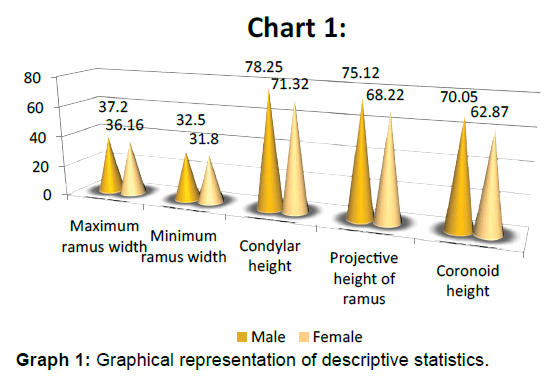
Out of the 500 digital orthopantomographs (OPGs) taken, 271 orthopantomographs (OPGs) were of females and 229 were of males. Descriptive statistics of mandibular rami measurements and all variables were found to be the best predictors for prediction of sex in the study with Variable C: Condylar height/ Maximum ramus height; Variable D: Projective height of ramus; and Variable E: Coronoid height, being highly significant, with p-values being <0.001. This implies that all variables showed their uniqueness among the individuals considered for the sample, with variables C: Condylar height/Maximum ramus height; Projective height of ramus; and E: Coronoid height, being highly significant, in their uniqueness. The associated univariate F-ratios for both the sexes are also shown in Table 1 and Graph 1.
| Variables | Sex | Wilks' Lambda | F-value | p-value | |||
|---|---|---|---|---|---|---|---|
| Male | Female | ||||||
| Mean | SD | Mean | SD | ||||
| Variable A | 37.20 | 3.88 | 36.16 | 3.54 | .981 | 9.789 | < 0.01 |
| Variable B | 32.50 | 3.14 | 31.80 | 3.32 | .989 | 5.726 | < 0.01 |
| Variable C | 78.25 | 5.09 | 71.32 | 5.06 | .683 | 231.463 | <0.001 |
| Variable D | 75.12 | 5.36 | 68.22 | 5.41 | .710 | 203.551 | <0.001 |
| Variable E | 70.05 | 5.77 | 62.87 | 4.94 | .689 | 224.500 | <0.001 |
** Highly significant at p<0.001
Table 1: Descriptive statistics
Also, it was observed that the mean values were significantly higher in males than the females for all variables. All the variables were found to be significant predictors for classifying a given sample based on sex. From the values obtained by linear discriminant function, calculations can be done with the help of the following equations to estimate sex in an unknown sample (Table 2).
| Variables | Sex | |
|---|---|---|
| Male | Female | |
| Variable A | .805 | .858 |
| Variable B | .538 | .666 |
| Variable C | 3.532 | 3.210 |
| Variable D | -1.294 | -1.159 |
| Variable E | .833 | .636 |
| (Constant) | -143.277 | -121.646 |
Table 2: Linear discriminant function
Males: - 143.277+0.805 (Maximum ramus width) +0.538 (Minimum ramus width) +3.532 (Condylar height) - 1.294 (Projective height of ramus) +0.833 (Coronoid height); and
Females: - 121.646+0.858 (Maximum ramus width) +0.666 (Minimum ramus width) +3.21 (Condylar height) - 1.159 (Projective height of ramus) +0.636 (Coronoid height).
For classifying a given sample as males or, females, the maximum value of two equations is considered. Prediction accuracy was calculated for the study sample. With all the variables in consideration, 80.4% of the sample was classified accurately (Table 3).
| Sex | Predicted Group | ||
|---|---|---|---|
| Male | Female | ||
| Original | Male | 174 | 55 |
| Female | 43 | 228 | |
| Male (%) | 76.0 | 24.0 | |
| Female (%) | 15.9 | 84.1 | |
80.4% of the original grouped cases were correctly classified
Table 3: Prediction accuracy
Among 271 females in the sample, 228 were estimated correctly as females using the above equations while in case of males, among 229 males in the study sample, 174 were estimated as males using the above equations with prediction accuracy being 84.1% amongst females, 76% amongst males and 80.4% for the entire study sample.
All the variables obtained were re-examined by two other expert Oral Radiologists to minimize inter-and intra-observer variability with the value of the Pearson’s correlation coefficient (r-value) being 0.98.
In the present study, sectioning point was found to be 0.19. For any unknown sample, for prediction of sex, we calculate the value obtained from the above mentioned equations using the 5 variables obtained from the Sidexis software. If the value is greater than this sectioning point i.e., 0.19, sample is male and if the value is lesser than this point that indicates a female (Table 4).
| Variables | Unstandardized Coefficients | Standardized Coefficients | Structure |
|---|---|---|---|
| Variable A | -.033 | -.120 | 0.175 |
| Variable B | -.080 | -.259 | 0.134 |
| Variable C | .201 | 1.021 | 0.851 |
| Variable D | -.084 | -.455 | 0.798 |
| Variable E | .123 | .656 | 0.839 |
| (Constant) | -13.315 |
Sectioning point: 0.19
Table 4: Standardized and unstandardized coefficients
Discussion
Forensic odontology is the study of dental applications in legal proceedings. Recent advances and current trends in forensic odontology include DNA profiling from teeth, palatal rugae and lip print patterns, novel methods of identification of human remains, innovative techniques in bite-mark analysis, contemporary methods for prediction of age and sex and the role of digital panoramic radiography in forensic odontology.
Forensic odontology plays a major role in identification in any sort of mass disaster events that result in multiple fatalities that may not be identified by means of conventional methods. One of the crucial aspects of forensic odontology is to predict sex using fragmented jaws as intact skull is not available for analysis in mass disasters [9,10].
Prediction of sex based on morphology is subjective and likely to be biased but methods based on measurements are accurate and can be used in determination of the same from skull based measurements that provide a more objective criteria to arrive-at such conclusions with a greater degree of accuracy, reliability and reproducibility [7,10,11].
Digital orthopantomographs (OPGs) have been widely used by the clinicians as an appropriate screening tool for the diagnosis of oral diseases. The major advantages of dental panoramic radiography include broad coverage, low patient radiation dose, ease of examination and a shorter time required to make images [12-14]. The inherent limitations of this technique, however, include magnification and geometric distortion, little alteration in vertical dimensions and the technique being sensitive to positioning errors [11,12]. In the present study, the same was used because of the inherent properties of contrast and brightness enhancement and enlargement of images which can provide an accurate and reproducible method of measuring the chosen points.
The selected variables of ramus were chosen because of less chances of alteration in these variables with advancing age, compared to measurements of the body of the mandible. Also, there are less geometric errors in the images obtained by digital ortho-pantomographs (OPGs) in the ramus region compared to midline structures of the mandible.
In the present study, mandibular rami measurements were subjected to discriminant functional analysis. All variables expressed strong sexual dimorphism with the mandibular rami with strongest univariate dimorphism in terms of condylar height, coronoid height followed by projective height of ramus and maximum and minimum ramus widths. The overall prediction rate using all the five variables was 80.4%.
Similar results were obtained by the studies conducted by Martin ES and HrdlicKa A who stated that measurements of the mandibular ramus height showed greater sexual dimorphism [15,16] Giles E concluded that the height of the symphysis, ramus and body, mandibular body length and bigonial diameter were useful measurements with greater sexual dimorphism [17].
Hanihara K did a study on Japanese crania and obtained a higher level of accuracy using measurements from the calvarium and mandible [18]. Loth and Henneberg, too, did a similar study on African and American samples and suggested that the degree of flexure of the posterior border of the ramus was as reliable for prediction of sex as the pelvis [19].
In another study conducted by Pokhrel and Bhatnagar on dry mandibles of Indian population, mandibular measurements were concluded to be significant predictors for sex in the defined population [20].
Few studies, however, have been conducted on population in South India, hence, the present study aimed at evaluating the reliability of mandibular rami in prediction of sex in the South Indian population and purpose the use of same in forensic analysis and anthropology.
DNA identification of mass disaster victims is the preferred technology due to its simplicity, adequate sensitivity, and high discrimination power, however, this cannot be considered as a whole and sole procedure in certain situations. If we consider the South Asian Tsunami as a group of smaller disasters affecting 12 different countries and producing victims from about 30 countries, the sheer scale of this unpredicted natural disaster with more than 200,000 victims is one of the principal challenges to face.
Furthermore, amongst the victims, a high number of relatives were expected in addition to entire families that died without any family reference to be compared with. The additional complication of reduced availability of direct reference samples remains a major factor to be considered in such situations. Also, this fact, further, gets complicated by the rate and speed of body recovery from the sea affecting DNA integrity in some cases. An undetermined proportion of bodies has not yet been discovered and might never be.
All these challenges require an approach to the identification process of the victims as an integral forensic science identification effort based not only upon DNA data but also, on forensic anthropology, fingerprints, odontology and radiology [21].
Conclusion
This study, thus, concludes with the observation that mandibular ramus can be included as one of the skeletal indicator for prediction of sex in forensic analysis and anthropology especially, in situations where the availability of soft tissues is questionable for analysis. The study found that rami measurements using digital orthopantomographs (OPGs) were reliable indicators in prediction of sex. Hence, the study suggests the use of mandibular rami in prediction of sex in forensic anthropology procedures.
However, since numerous studies conducted in the past have demonstrated that skeletal characteristics differ in each population and have emphasized the need for population-specific osteometric standards for sex determination, the present study, also, paves way for further studies to be conducted on more diverse populations to establish population-specific osteometric standards and deriving appropriate regression equations taking into consideration numerous factors including the various socioenvironmental factors e.g. nutrition, food, climate; pathologies influencing the development and thus, the appearance of bones and other factors which might influence the growth patterns to come to valid conclusions.
References
- Patil KR, Mody RN. Determination of sex by discriminant function analysis and stature by regression analysis: A lateral cephalometric study. Forensic Science International. 2005; 147:175-80.
- World Disasters Report 2012. International Federation of Red Cross and Red Cross Societies.
- Saini V, Srivastava R, Shamal SN, Singh TB, Pandey AK, Tripathi SK. Sex determination using mandibular ramus flexure: A preliminary study on Indian population. Journal of Forensic and Legal Medicine. 2011; 18:208-12.
- Iván Claudio Suazo Galdames, Jaime San Pedro Valenzuela, Nilo Alejandro Schilling Quezada, César Eduardo Celis Contreras, José Alejandro Hidalgo Rivas, Mario Cantín López. Ortopantomographic blind test of mandibular ramus flexure as a morphological indicator of sex in Chilean young adults. International Journal of Morphology 2001; 26:89-92.
- Rosing FW, Graw M, Marre B, Ritz-Timme S, Rothschild MA, et al. Recommendations for the forensic diagnosis of sex and age from skeletons. Homo 2007; 58:75-89.
- Maryna S, Yasar IM. Sexual dimorphism in the crania and mandibles of South African whites. Forensic Science International 1998; 98:9-16.
- Humphrey LT, Dean MC, Stringer CB. Morphological variation in great ape and modern human mandibles. Journal of Anatomy 1999; 195:491-513.
- Kumar MP, Lokanadham S. Sex determination and morphometric parameters of human mandible. International Journal of Research in Medical Sciences. 2013; 1:93-96.
- Kahana T, Hiss J. Forensic radiology. The British Journal of Radiology. 1999; 72:129-133.
- Indira AP, Markande A, David MP. Mandibular ramus, an indicator for sex determination: A digital radiographic study. Journal of Forensic Dental Sciences. 2012; 4:58-62.
- Press SJ, S Wilson. Choosing between logistic regression and discriminant analysis. Journal of the American Statistical Association. 1978; 73:699-705.
- Tahmine Razi, Seyed Hosein Moslemzade, Sedighe Razi. Comparison of Linear Dimensions and Angular Measurements on Panoramic Images Taken with Two Machines. Journal of Dental Research, Dental Clinics and Dental Prospects. 2009; 3:7-10.
- Laster WS, Ludlow JB, Bailey LJ, Garland Hershey H. Accuracy of measurements of mandibular anatomy and prediction of asymmetry in panoramic radiographic images. Dentomaxillofacial Radiology. 2005; 34:343-9.
- Schulze, Krummenauer F, Schalldach F, d'Hoedt B. Precision and accuracy of measurements in digital panoramic radiography. Dentomaxillofacial Radiology. 2000; 29:52-6.
- Martin ES. A study of an Egyptian series of mandibles with special reference to mathematical methods of sexing. Biometrika. 1936; 28:149-78.
- HrdlicKa A. Mandibular and maxillary hyperostoses. American Journal of Physical Anthropology. 1940; 27:1-55.
- Giles E. Sex determination of discriminant function analysis of the mandible. American Journal of Physical Anthropology. 1964; 22:129-35.
- Hanihara K. Sex diagnosis of Japanese skulls and scapulae by means of discriminant function. Journal of the Anthropological Society of Nippon. 1959; 67:191-7.
- Loth SR, Henneberg M. Mandibular ramus flexure: New morphologic indicator of sexual dimorphism in the human skeleton. American Journal of Physical Anthropology. 1996; 99:473-486.
- Pokhrel R, Bhatnagar R. Sexing of mandible using ramus and condyle in Indian population: A discriminant function analysis. European Journal of Anatomy. 2013; 17:39-42.
- Fleck F. Tsunami body count is not a ghoulish num


 The Annals of Medical and Health Sciences Research is a monthly multidisciplinary medical journal.