Applications of Stem Cells in Interdisciplinary Dentistry and Beyond: An Overview
- *Corresponding Author:
- Dr. Shalu Rai
Department of Oral Medicine and Radiology, Institute of Dental Studies and Technologies, Kadrabad, Modi Nagar, Uttar Pradesh - 201 201, India.
E-mail: drshalurai@gmail.com
Abstract
In medicine stem cell–based treatments are being used in conditions like Parkinson’s disease, neural degeneration following brain injury, cardiovascular diseases, diabetes, and autoimmune diseases. In dentistry, recent exciting discoveries have isolated dental stem cells from the pulp of the deciduous and permanent teeth, from the periodontal ligament, and an associated healthy tooth structure, to cure a number of diseases. The aim of the study was to review the applications of stem cells in various fields of dentistry, with emphasis on its banking, and to understand how dental stem cells can be used for regeneration of oral and non‑oral tissues conversely. A Medline search was done including the international literature published between 1989 and 2011. It was restricted to English language articles and published work of past researchers including in vitro and in vivo studies. Google search on dental stem cell banking was also done. Our understanding of mesenchymal stem cells (MSC) in the tissue engineering of systemic, dental, oral, and craniofacial structures has advanced tremendously. Dental professionals have the opportunity to make their patients aware of these new sources of stem cells that can be stored for future use, as new therapies are developed for a range of diseases and injuries. Recent findings and scientific research articles support the use of MSC autologously within teeth and other accessible tissue harvested from oral cavity without immunorejection. A future development of the application of stem cells in interdisciplinary dentistry requires a comprehensive research program.
Keywords
Banking, Dental stem cells, Interdisciplinary dentistry
Introduction
Stem cells are immature, undifferentiated cells that can divide and multiply for an extended period of time, differentiating into specific types of cells and tissues.[1] They are defined as cells that self-replicate and are able to differentiate into at least two different cell types. Both criteria must be present for a cell to be called a ‘stem cell’.[2] Discoveries in stem cell research present an opportunity for scientific evidence that stem cells, whether derived from adult tissues or the earliest cellular forms, hold great promise that goes far beyond regenerative medicine. Dentists are at the forefront of engaging their patients in potentially life-saving therapies derived from their own stem cells located either in deciduous or permanent teeth.
In 2000, the National Institute of Health mentioned the discovery of adult stem cells in the impacted third molars and even more resilient stem cells in the deciduous teeth, thus providing the prospect of regeneration of dentin and/or dental pulp; biologically viable scaffolds will be used for replacement of the orofacial bone and cartilage and defective salivary glands, which can be partially or completely regenerated.[1]
Tooth banking is based on the firm belief that personalized medicine is the most promising avenue for treating challenging diseases and injuries that would occur throughout life. Individuals have different opportunities at different stages of their life for banking their valuable cells. Recent studies have shown that stem cells from human exfoliated deciduous teeth (SHED) have a greater ability to develop into various types of body tissues compared to other types of stem cells.
Methods of Literature Search
The literature was searched using Pubmed and electronic data bases from 1999 to 2011. Key words such as stem cells, banking, SHED, and regeneration were used. The search was restricted to English language articles, published from 1999 to 2011, and various in vivo and in vitro studies were included. The purpose of this review was to highlight the application of stem cells in various fields of dentistry, with emphasis on its banking.
The principle stem cells are of two types:
Embryonic stem cells
They are derived from cells of the inner cell mass of the blastocysts, during embryonic development. They are pluripotent, with the blastocyst being the early stage embryo consisting of approximately 50-150 cells, and give rise to all derivatives of three primary germ layers, that is, ectoderm, endoderm, and mesoderm. When given sufficient and necessary stimulation for a specific cell type, they can develop into more than 200 cell types of the adult body, but they do not contribute to the extra embryonic membrane or placenta. The most important and potential use of embryonic stem cells (ESC), is clinically in transplantation medicine, where they can be used to develop cell replacement therapies.[1-4]
The FDA-approved human trials using ESC for treatment of paralysis from a spinal cord injury[5] and diseases of the eye have been reported.[6]
Adult stem cells
Adult stem cells are multipotent, because their potential is normally limited to one or more lineages of specialized cells.[3] They are not subject to the ethical controversy that is associated with ESC.[1]
The adult stem cells can be recovered from the following:
1. Bone marrow–derived mesenchymal stem cells: Bone marrow transplants were the first successful stem cell therapies. Presently peripheral blood stem cell collection is being used in place of bone marrow aspiration.[7]
2. Adipose-derived adult stem cells: Have also been isolated from human fat, usually by the method of liposuction.[8]
3. Umbilical cord stem cells: Are derived from the blood of the umbilical cord.[9,10]
4. Amniotic fluid–derived stem cells: Can be isolated from the aspirates of amniocentesis during genetic screening or collection at the time of delivery.[11]
5. Induced pluripotent stem cells: Refer to adult or somatic stem cells that have been genetically reprogramed to behave like ESC. Takahashi (2006)[12] reported a generation of iPS from mouse embryonic fibroblasts and adult mouse tail-tip fibroblasts by the retrovirus-mediated transfection of four transcription factors, namely Oct3/4, Sox2, c-Myc, and Klf4, but it was Oct 4, Sox2, Nanog, and Lin28 according to Yu, et al. (2007).[13] Takahashi (2007)[14] demonstrated the generation of iP cells from adult human dermal fibroblasts with the same four factors: Oct3/4, Sox2, Klf4, and c-Myc. Human iPS cells were similar to human embryonic stem (ES) cells in morphology, proliferation, surface antigens, gene expression, epigenetic status of pluripotent cell-specific genes, and telomerase activity. Furthermore, these cells expressed stem cell markers and are capable of generating cells characteristic of all three germ layers.
Successful reprograming of differentiated human somatic cells into a pluripotent state would allow the creation of patient-and disease-specific stem cells, thus showing the capacity to generate a large quantity of stem cells as an autologous cell source, which can be used to regenerate patient-specific tissues and they also appear to minimize the need for ES cells.
Various techniques have been used to alter gene expression in stem cells, which include, cell transfection via electroporation, lipid-mediated delivery or biolistics particle delivery, as well as cell transduction through viral-mediated gene delivery.[15]
Transfection and nucleofection methods have been tried in the laboratory using plasmids, with an enhanced green fluorescence protein (eGFP) as a reporter.[16]
6. Dental stem cells: Are the most accessible stem cells. They are isolated from the dental pulp of healthy teeth, both primary and permanent teeth, the periodontal ligament, including the apical region of developing teeth, and other tooth structures. Craniofacial stem cells, including dental stem cells (DSC), originate from neural crest cells and mesenchymal cells during development.[17]
Two major cell types are involved in dental hard tissue formation: Epithelium - derived ameloblasts that form enamel and the mesenchymal - originated odontoblasts that is responsible for the production of dentin.[18]
Epithelium stem cells
Although significant progress has been made with mesenchymal stem cells, there is no information available on the use of Epithelium stem cells in humans, because their ameloblasts and ameloblast precursors are eliminated soon after eruption.[18] Stem cell technology appears to be the only possibility to re-create an enamel surface.[18,19]
Mesenchymal stem cells
Mesenchymal stem cells (MSC) possess a high self-renewal capacity and the potential to differentiate into mesodermal lineages, thus forming cartilage, bone, adipose tissue, and skeletal muscle, and participate in the formation of many craniofacial structures.[20] It can be used autologously without concern of immunorejection, as it can be isolated from patients who need the treatment.[1] MSC have been used allogenically to heal large defects.[2,21]
The following dental mesenchymal progenitors have been used for tooth engineering purposes:[4,22,23]
SHED
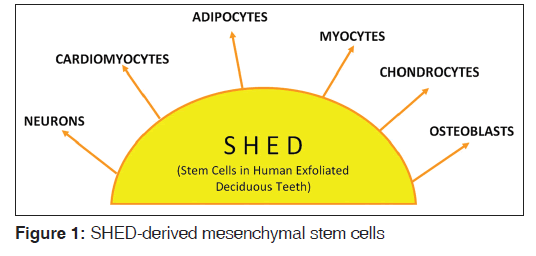
These cells are immature, unspecialized cells in the teeth that are able to grow into specialized cell types by a process called ‘differentiation’. In 2003, Miura et al., isolated cells from the deciduous dental pulp, which were highly proliferative and clonogenic.[24] Abbas et al., 2008,[19] investigated that SHED Teeth were of neural crest origin. SHEDs were found to express early mesenchymal stem cell markers (STRO-1 and CD146).[25] These cells exhibited high plasticity, as they could differentiate into adipocytes,[18,24,26,27] chondontocytes, osteoblasts,[24,28,29] and neurons in vitro.[26,27,30,31] [Figure 1]. After in vivo implantation, the SHED cells could induce bone or dentin formation, but in contrast to dental pulp stem cells (DPSC), they failed to produce a dentin-pulp complex and represented an immature population of multipotent stem cells.[18,24,27]
Adult DPSCs
Adult DPSCs are isolated from adult dental pulp[1,2,32] and contain precursors capable of forming odontoblasts under appropriate signals like calcium hydroxide or calcium phosphate materials, which contain pulp capping materials used by a dentist for treatment. The in vivo therapeutic targeting of these adult stem cells remains to be explored.[2]
Periodontal ligament stem cells
Periodontal ligament stem cells (PDLSCs) have a multilineage differentiation potential and are able to undergo adipogenic, osteogenic, and chondrogenic phenotype in vivo.[26] PDLSCs are isolated from the separated PDL of the roots of the impacted human third molar and are found to express STRO-1 and CD146.[16,27] Seo, et al.,[33] 2004, has demonstrated that PDL itself contains progenitors that can be activated to self-renew and regenerate other tissues like cementum and alveolar bone.[18,32] However, they formed sparse calcified nodules compared to DPSCs.[4]
Stem cells from the apical part of the papilla
In 2006, Sonoyama et al.,[34] isolated a new population of dental stem cells, and called them stem cells from the apical part of the papilla (SCAPs). SCAPs are clonogenic fibroblast-like cells, but have a higher proliferation rate than DPSCs. Similar to other dental stem cells, SCAPs express the early mesenchymal surface markers, STRO-1 and CD146.[27]
As in the case of DPSCs, when SCAPs were transplanted into immunocompromised mice in an appropriate carrier matrix, a typical dentin pulp–like structure was formed, with odontoblast-like cells.[18,27]
Stem cells from the dental follicle
In 2005, Morsczeck et al.,[35] isolated Stem Cells from the dental follicle of human third molars, which expressed the stem cell markers Notch1, STRO-1, and nestin.[18,27] An in vitro study demonstrated the potential of DFPCs to undergo osteogenic, adipogenic, and neurogenic differentiation.[27] After in vivo implantation, the immortalized dental follicle cells were able to recreate a new periodontal ligament (PDL).[36]
Bone marrow–derived mesenchymal stem cells
Bone marrow-derived mesenchymal stem cells (BMSC) originate from the mesoderm.[18] They can be obtained from several other sources such as the synovial membrane[37] and periosteum,[38] but it remains to be explored as to which source can be used for optimal tooth development, for clinical application. BMSC are able to form in vivo cementum, PDL, and alveolar bone, after implantation into defective periodontal tissues. Thus, they provide an alternative source of MSC for the treatment of periodontal diseases.[39]
Tooth stem cells have an advantage over ESC in treating diseases, because they are less likely to develop into teratomas (tumors) when transplanted. However, the limitation of using DSC is that it is difficult to harvest a large quantity of stem cells from the teeth, the requirement of a professional technician is higher for extraction, isolation, and culture, and it also takes a longer time to culture mesenchymal stem cells from the teeth-active tissue.[40]
Banking of dental stem cells
The key to successful stem cell therapy is to harvest cells and store them safely until accident or disease requires their usage. Tooth banking is not very popular, but the trend is catching up, mainly in the developed countries.[26]
In the year 2003, Dr. Songtao Shi, a pediatric dentist, was able to isolate, grow, and preserve tooth stem cell’s regenerative ability, by using the deciduous teeth of his six-year-old daughter.[24] He called the cells as SHED. The existing research has shown that primary teeth are a better source of therapeutic stem cells for use in regenerative medicine than wisdom teeth, and orthodontically extracted teeth.[29]
Advantages of SHED banking
1. Provides an autologous transplant for life
2. Simple and painless procedure[24]
3. SHED cells are complementary to stem cells from the cord blood[29]
4. Useful for close relatives of the donor.[3]
5. Not subjected to the same ethical concerns as embryonic stem cells.[2,3]
Collection, Isolation, and Preservation
For deciduous teeth, the best candidates for isolation are canines and incisors with the presence of healthy pulp that is starting to loosen. Adolescents have two excellent opportunities for banking, following extraction of bicuspid teeth for orthodontic treatment and at the time of extraction of wisdom teeth. The follicular sac of an unerupted tooth is also a valuable source for stem cells.[3,26]
Step 1: Tooth collection
First step is to place the tooth in sterile saline solution,[26] or in fresh milk, in a storage container along with frozen gel packs, in a Kit, which is then ready for delivery to the laboratory.[41] The exfoliated tooth should have a pulp red in color, indicating that the pulp has received blood flow up until the time of removal, which is indicative of cell viability. The gray color of the pulp indicates that the blood flow is compromised, and thus the stem cells are likely to be necrotic and are no longer viable for recovery. With the recovery of the tooth it is transferred into a vial containing hypotonic phosphate buffered saline solution, which helps to prevent the tissue from drying during transport (up to four teeth in one vial). The vial is then carefully sealed and placed into a thermette (a temperature phase change carrier), which is then placed into an insulated metal transport vessel. This procedure maintains the sample in a hypothermic state during transportation, which is known as ‘sustentation’.[26]
Step 2: Stem cell isolation
When the tooth bank receives the Kit or vial, all the cells are isolated and a stringent protocol is followed for cleaning the tooth surface by various disinfectants. The pulp tissue is isolated from the pulp chamber and the cells are then cultured in a MSC medium, under appropriate conditions.[24,26] By making changes in the MSC medium different cell lines can be obtained such as odontogenic, adipogenic, and neural. If contamination is extensive, then a change in procedures can be performed using STRO 1 or CD 146.[26] The time from harvesting to arrival at the processing storage facility should not exceed 40 hours.
Step 3: Stem cell storage
Either of the following approaches can be used.
a. Cryopreservation:[26,41,42,43] cells are preserved in liquid nitrogen vapor at −150º thus maintaining their latency and potency so that the cells can be stored for a long time and still remain viable for use.
b. Magnetic freezing:[26,44] or the Cell Alive System (CAS), uses a magnetic field, where the object is chilled below freezing point, which ensures distributed low temperature without damaging the cell wall. The Hiroshima University claims that this can increase the cell survival rate in teeth to a high of 83%.
Licensed tooth stem cell banks, Internationally and in India, used for cryopreservation and isolation are as follows:[26,45]
1. In Japan, the first tooth bank was established in Hiroshima University and the company was named as ‘Three Brackets’ (Suri Buraketto).
2. BioEden (Austin, Texas), StemSave, and Store-a-Tooth (USA)
3. The norwegian tooth bank
4. In India, Stemade Biotech Pvt. Ltd. (Delhi, Chennai, Chandigarh, Pune, and Hyderabad).
Tooth regeneration
Langer and Vacanti first described tissue engineering as, ‘an interdisciplinary field that applies the principles of engineering and life sciences toward the development of biological substitutes that restore, maintain, or improve tissue function’.[46] Although there can be many differing definitions of regenerative medicine, in practice, the term has come to represent applications that repair or replace structural and functional tissues, including bone, cartilage, and blood vessels, among organs and tissues.[47] Tissue engineering is based on the principles of molecular developmental biology governed by bioengineering.[46,48-50] The three main components for morphogenesis and tissue engineering are: Inductive signals, responding cells, and a scaffold.[49,50-53]
Currently there are two major approaches to tooth regeneration.[54]
1. By using the principles of tissue engineering
2. By reproducing the developing processes of embryonic tooth formation
The Yelick group used dental epithelium and mesenchymal tissues to seed heterogenous single cells onto a scaffold-like polycoglycolic polymer resulting in tiny tooth-like tissues (enamel, dentin, and pulp), but further studies are required to achieve reconstituted and structurally sound teeth.[55]
Hu, et al.,[56] have shown that BMC can give rise to ameloblast-like cells, while Sharpe’s, et al.,[57] reported that BMC, when placed in contact with the oral epithelium, can differentiate into dental mesenchymal cells, that is, dentin and pulp.
Sonoyama, et al.[34] and Huang, et al.,[58] explored that in miniature pigs, the bio-root periodontal complex is built up by postnatal stem cells using SCAP and PDLSC, to which an artificial porcelain crown is fixed, and concluded that dental stem cells engineering can be used for tooth root regeneration.
Ikeda, et al.,[59] explored in adult mouse that fully functioning tooth replacement can be achieved by transplantation of a bioengineered tooth germ into an alveolar bone of a lost tooth region by the interaction between the dental epithelium and mesenchymal cells. The bioengineered tooth, which had erupted and reached occlusion, had the correct tooth structure and hardness of mineralized tissues for mastication.
As suggested by Nakahara and Idle,[60] there are few limitations in tooth regeneration.
Principles of tissue engineering, related to tooth regeneration, which might not mimic tooth morphology.
At the present time there is no embryonic environment that enables bone marrow cells to differentiate into tooth germ cells.
Also, host immune rejection and ethical issue on the use of human embryo still exist to date.
Application in Interdisciplinary Dentistry
Uses in endodontics
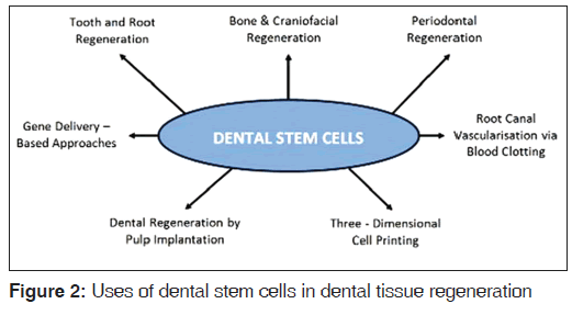
Based on the basic tissue engineering principles,[61-64] Peter Murray et al.,[61] identified several major areas of research that might have applications in the development of these techniques, which are: [Figure 2]
Concept of root canal revascularization via blood clotting
Several case reports have documented revascularization[65,66] of the necrotic root canal systems by disinfection followed by establishing bleeding into the canal system[58] via overinstrumentation.[67-69] Use of intracanal irrigants (NaOCl and chlorhexdine) along with the placement of antibiotics (e.g., a mixture of ciprofloxacin, metronidazole, and minocycline paste), for several weeks, is a critical step, as it effectively disinfects the root canal systems and increases revascularization of the avulsed and necrotic teeth. The revascularization process offers negligible chances of immune rejection and pathogen transmission, as regeneration of the tissue takes place by the patient’s own blood cells. However, some critical limitations of this technique entail that caution is required, as the source of regenerated tissue has not been identified and also the concentration and composition of cells trapped in the fibrin clot are unpredictable.[61] Animal and more clinical studies are required, to investigate the potential of this technique, before it can be recommended for general use in patients.
Postnatal stem cell therapy
The process comprises of postnatal stem cells (derived from skin, buccal mucosa, fat, and bone)[70] being injected into disinfected root canal systems after the apex is opened. This process has many advantages like the harvesting and delivery of autogenous stem cells by syringe, being relatively easy; and the potential of these cells to induce new pulp regeneration.[61] However, there are several disadvantages, like the cells may have a low survival rate and they may migrate to different locations within the body.[71] Instead, all three elements (cells, growth factors, and scaffold) must be considered, to maximize the potential for success of pulp regeneration.[61,72]
Pulp implantation
The pulp cells can be grown on biodegradable membrane filters to transform two-dimensional into three-dimensional cell cultures.[73] The ease of growing these cells on filters in the laboratory, for evaluation of cytotoxicity of test materials, is recognized as the main advantage of this delivery system. The potential problems associated with the implantation of sheets of cultured pulp tissue is that it requires specialized procedures for proper adherance to the root canal walls. As sheets of cells lack vascularity, only the apical portion of the canal systems will receive these cellular constructs, with coronal canal systems filled with scaffolds capable of supporting cellular proliferation.[61]
Scaffold implantation and delivery
A scaffold should contain growth factors, Bone Morphogenic Protein (BMP), fibroblast growth factors, and Vascular endothelial growth factors,[74] to aid stem cell proliferation and differentiation, apart from having nutrients promoting cell survival and growth as well as antibiotics to prevent any bacterial in-growth in the canal systems. The scaffold materials may be natural or synthetic, biodegradable or permanent. The synthetic materials like polylactic acid, polyglycolic acid, and polycaprolactone degrade within the human body and have been succesfully used for tissue engineering purposes.[75] Limitations consist of difficulties of obtaining high porosity and regular pore size.[76]
Hydrogels are injectable scaffolds that can be delivered by syringe[77,78] and have the potential to be noninvasive and easy to deliver into root canal systems. Despite these advances they are at an early stage of research. To make hydrogels more practical, research is focusing on making them photopolymerizable to form rigid structures once they are implanted into the tissue site.[79]
Three-dimensional cell printing
The three-dimensional cell printing technique can be used to precisely position cells so that they have the potential to create tissue constructs that mimic the natural tooth pulp tissue structure [Figure 2].[80]
Careful orientation of the pulp tissue construct during placement into the cleaned and shaped root canal systems in accordance with its apical and coronal asymmetry is the prime requisite for the success of the technique. However, early research has yet to show that three-dimensional cell printing can create functional tissue in vivo.[61,81]
Gene therapy
This technique involves a gene encoding a therapeutic protein being introduced into the cells, which can then express the target protein.[82-86] A recent review has discussed the use of gene delivery in regenerative endodontics.[49] Rutherford,[87] in his study, used ferret pulps with cDNA (complementary DNA)-transfected mouse BMP-7, but failed to produce a reparative response, suggesting further research in the potential of pulp gene therapy. Although viral delivery systems have been used successfully in a broad range of tissues, they have serious health hazards, including the risks for mutagenesis, carcinogenesis, and invoking immune reactions in response to viral infection or viral proteins.[61] At this time the potential benefits and disadvantages are largely therotical.[61,88]
Huang, et al. explored in mice that pulp-like tissue can be regenerated de novo in an emptied root canal space by stem cells from apical papilla and dental pulp that give rise to odontoblast-like cells, producing dentin-like tissue on the existing dentinal walls via stem/progenitor cell-based approaches and tissue engineering technologies.[89]
Uses in periodontology
The use of growth and differentiation factors for regenerating periodontal tissue is the most popular tissue engineering approach. To date several growth factors including Transfoming growth factors-b (TGF-b) superfamily members, such as, bone morphogenetic protein-2 (BMP-2), BMP-6, BMP-7, BMP-12, TGF-b, basic fibroblast growth factors (bFGF), and platelet derived growth factors (PDGF), have been used as a protein-based approach to regenerate periodontal tissues.[90-97] Several preclinical studies using MSCs have shown efficient reconstruction of bone defects larger than those that would spontaneously heal.[1,21] Kramer, et al.,[98] demonstrated that PDL-like tissue can be developed from periodontal progenitor cells and from mesenchymal stem cells in contact with either PDL factors or the tissue itself. Kawaguchi and colleagues 2004,[38] used bone marrow−derived MSCs in combination with atelocollagen for regeneration of periodontal tissues in class III furcation defects, in dogs, and concluded that additional studies would be required using different scaffold materials and a various range of cell concentration to obtain conclusive results.[82] Although encouraging results for periodontal tissue regeneration have been found in numerous clinical investigations, limitation xists, with topical protein delivery. Dual growth factor delivery technologies could be useful in periodontal tissue engineering.[82]
Uses in oral maxillofacial surgery for craniofacial reconstruction
Stem cells have been used in the tissue engineering of a human-shaped temporomandibular joint.[1,4,99] Alhadlaq and Mao,[21,100] used MSC-derived cells encapsulated in a poly {ethylene glycol} diacrylate hydrogel that was molded into an adult human mandibular condyle in stratified yet integrated layers of cartilage and bone. The osteochondral grafts, in the shape of human TMJs, were implanted in immunodeficient mice for up to 12 weeks. Upon harvest, the tissue-engineered mandibular joint condyles retained their shape and dimensions.[1]
According to Pittenger, et al.,[101,102] bone marrow–derived MSC are now under consideration for the repair of craniofacial bone and even the replacement or regeneration of oral tissues.[1,103] Reconstruction of craniofacial and dental defects using MSC avoids many of the limitations of both auto- and allografting techniques.[99] Clinical studies are being conducted using stem cells for alveolar ridge augmentation and long-bone defects.[103,104] Vascularized bone grafts are also used in development using stem cells, and reconstruction of a patient’s resected mandible has been carried out using this technique [Figure 2].[105]
Changes in the expression of stem cell markers in oral mucosal lesions and oral squamous cell c100arcinoma
Despite the pivotal role of stem cells in the homeostasis of oral epithelium, the location of this cell population within the tissue is uncertain. How disease influences these cells in vivo also remains to be elucidated.
Kose, et al.,[106] 2007, in a study, described the expression of six putative stem cell markers, that is, a 6 and b 1 integrins, melanoma-associated chondroitin sulfate proteoglycan (MCSP), NG2 the rat homolog of human MCSP, notch 1, and keratin 15 (k15), in normal buccal epithelium and showed their expression to be altered in oral mucosal diseases, oral lichen planus, and oral hyperkeratotic lesions. This suggest that stem cell marker expression may be altered by pathological signaling in these lesions. Further studies of these molecular perturbations are essential to understand the fundamental role of adult stem cells in the pathogenesis of benign mucosal diseases.
Chiou, et al.,[107] in his study, suggests that identification of a rare subpopulation of cancer cells, termed cancer stemcells, from oral squamous cell carcinoma (OSCC) facilitates the monitoring, therapy, or prevention of OSCC. Enriched oral cancer stem-like cells (OC-SLC) expressed stem/progenitor cell markers greatly, as also the ABC transporter genes (Oct-4, Nanog, CD117, Nestin, CD133, and ABCG2), which displayed enhanced migration/ invasion/malignancy capabilities in vitro and in vivo. The author concluded that Nanog/Oct-4/CD133 triple-positive patients predicted the worst survival prognosis of OSCC patients.
Application of dental stem cell for regeneration of non-dental tissues
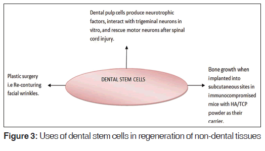
Fang, et al., 2007,[108] demonstrated that PDLSCs could form substantial amounts of collagen fibers and improved facial wrinkles in mice. Otaki, et al.,[109] 2007, reported that dental pulp cells produced bone when implanted into subcutaneous sites in immunocompromised mice with HA/TCP (Hydroxy apatite/Tricalcium phosphate) powder as a carrier. Nosrat, et al., 2001,[110] reported grafting dental pulp tissues into hemisected spinal cord increases the number of surviving motoneurons, indicating a functional bioactivity of the dental pulp–derived neurotrophic factors, rescuing motoneurons in vivo [Figure 3].
Conclusion
The current research on dental stem cells is expanding at an unprecedented rate. MSC–derived chondrocytes can be used for reconstruction of orofacial cartilage structures such as temporomandibular joint and nasal cartilage. MSC–derived osteoblast can be used for regeneration of oral and craniofacial bones. MSC–derived myocytes can be used to treat muscular dystrophy and facial muscle atrophy. MSC–derived adipocytes have the ability to repair damaged cardiac tissues following a heart attack. It is now possible to cryopreserve healthy teeth as sources of autogenous stem cells, either when they are exfoliated or by extraction, because the opportunity to bank a patient’s dental stem cells will have the greatest future impact if seized when patients are young and healthy, for future regenerative therapies. The conservative treatment of life-threatening and disfiguring defects and diseases and the ability to treat currently incurable diseases are becoming a reality. To date, the research is only confined to animal models and more human research trials are needed to determine the therapeutic utility of stem cells. Stem cell costs, coverage by health insurance, and role of pharmaceutical companies, play a pivot role in determining the future of stem cells. Some applications are approved or are being reviewed by the FDA, in the United States, and equivalent regulatory agencies elsewhere.
Source of Support
Nil.
Conflict of Interest
None declared.
References
- Mao JJ, Collins FM. Stem Cells: Sources, therapies and the dental professional. Available from: http://www.ineedce. com/courses/1486/PDF/StemCells.pdf. [Last cited on 2011 May 10].
- Mao JJ. Stem cell and future of dental care. NY State Dent J 2008;74:20-4.
- Reznick JB. Stem Cells: Emerging medical and dental therapies for the dental professional. Oct. Available from http://www. stemsave.com/Docs/News/Dentaltown%20StemCell%20 CE.pdf. 2008. [Last cited on 2011 May 2].
- Nedel F, André Dde A, de Oliveira IO, Cordeiro MM, Casagrande L, Tarquinio SB, et al. Stem cells: Therapeutic potential in dentistry. J Contemp Dent Pract 2009;10:90-6.
- Severine Kirchner S. “Arise and Walk!” – A miracle cure in the works. Available from: http://dailyreckoning.com/ arise-and-walk-a-miracle-cure-in-the-works. [Last accessed on 2012 Feb 27].
- Walsh. F First trial results of human embryonic stem cells. Available from: http://www.bbc.co.uk/news/ health-16700394. [Last accessed on 2012 Feb 27].
- Pompilio G, Cannata A, Peccaori F, Bertolini F, Nascimbene A, Capogrossi MC, et al. Autologous peripheral blood stem cell transplantation for myocardial regneneration. A novel stratergy for cell collection and surgical injection. Ann Thorac Surg 2004;78:1808-12.
- De Ugarte DA, Morizona K, Elbarbary A, Alfonso Z, Zuk PA, et al. Comparison of multilineage cells from human adiposetissue and bone marrow. Cells Tissue Organ 2003;174:101-9.
- Laughlin MJ. Umblical cord blood for allogenic transplantation in children and adults. Bone Marrow Transplant 2001;27:1-6.
- Gang EJ, Jeong JA, Hong SH, Hwang SH, Kim SW, Yang IH, et al. Skeletal myogenic differentiation of mesenchymalstem cells isolated from human umbilical cord. Stem cells 2004;22:617-24.
- De Gemmis P, Lapucci C, Bertelli M, Tognetto A, Fanin E, Vettor R, et al. A real–time PCR approach to evaluate adipogenic potential of amniotic fluid-derived human mesenchymal stem cells. Stem Cells Dev 2006;15:719-28.
- Takahashi K, Yamanaka S. Induction of pluripotent stem cells from mouse embryonic and adult fibroblast cultures by defined factors. Cell 2006;126:663-76.
- Yu J, Vodyanik MA, Smuga-Otto K, Antosiewicz-Bourget J, Frane JL, Tian S, et al. Thomson. Induced pluripotent stem cell lines derived from human somatic cells. Science 2007;21:1917-20.
- Takahashi K, Tanabe K, Ohnuki M, Narita M, Ichisaka T, Tomoda K, et al. Induction of pluripotent stem cells from adult human fibroblast by defined factors. Cell 2007;131:861-72.
- Transfection of Stem Cells. Available from: http://www. bio-rad.com/evportal/en/US/evolutionPortal.portal?_ nfpb=true and _pageLabel=SolutionsLandingPage and catID=LUSR3BMNI. [Last accessed on 2012 Feb 27]
- Chatterjee P, Cheung Y, Liew C. Transfecting and nucleofecting human induced pluripotent stem cells. J Vis Exp 2011;pii: 3110.
- Caplan AI. Mesenchymal stem cells. J Orthop Res 1991;9:641-50.
- Bluteau G, Luder HU, De Bari C, Mitsiadis TA. Stem cells for tooth engineering. Eur Cell Mater 2008;16:1-9.
- Yelick PC, Vacanti JP. Bioengineered teeth from tooth bud cells. Dent Clin N Am 2006;50:191-203.
- Bongso A, Lee EH. Stem cells: From bench to bedside. 2nd ed. Singapore: World Scientific; 2005.
- Alhadlaq A, Mao JJ. Mesenchymal stem cells: Isolation and therapeutics. Stem Cells Dev 2004;13:436-48.
- Peneva M, Mitev V, Ishketiev N. Isolation of mesenchymalstem cells from the pulp of deciduous teeth. Journal of IMAB-Annual Proceeding (Scientific Papers) 2008, book 2. Available from http://www.journal-imab-bg.org/statii-08/vol08_2_84-87str.pdf.
- D’Aquino R, De Rosa A, Laino G, Caruso F, Guida L, Rullo R, et al. Human dental pulp stem cells: From biology to clinicalapplications. J Exp Zool B Mol Dev Evol 2009;312B: 408-15.
- Miura M, Gronthos S, Zhao M, Lu B, Fisher LW, Robey PG, et al. SHED: Stem cells from human exfoliated deciduousteeth. Proc Natl Acad Sci USA 2003;100:5807-12.
- Abbas, Diakonov I, Sharpe P. Neural Crest Origin of Dental Stem Cells. Pan european federation of the international association for dental research (PEF IADR). Seq #96-Oral Stem Cells: Abs, 0917, 2008.Available from http://iadr.confex. com/iadr/pef08/techprogram/abstract_111214.htm.
- Arora V, Arora P, Munshi AK. Banking Stem cells from human exfoliated deciduous teeth (SHED): Saving for the future. J Clin Pediatr Dent 2009;33:289-94.
- Jamal M, Chogle S, Goodis H, Karam SM. Dental stem cells and their potential role in regenerative medicine. J Med Sci 2011;4:53-61.
- de Mendonça Costa A, Bueno DF, Martins MT, Kerkis I, Kerkis A, Fanganiello RD, et al. Reconstruction of large cranial defects in nonimmunosuppressed experimental design with human dental pulp stem cells. J Craniofac Surg 2008;19:204-10.
- Shi S, Bartold PM, Miura M, Seo BM, Robey PG, Gronthos S. The efficacy of mesenchymal stem cells to regenerate and repair dental structures. Orthod Craniofac Res 2005;8:191-9.
- Arthur A, Rychkov G, Shi S, Koblar SA, Gronthos S. Adult human dental pulp stem cells differentiate toward functionally active neurons under appropriate environmental cues. Stem Cells 2008;26:1787-95.
- Kerkis I, Ambrosio CE, Kerkis A, Martins DS, Zucconi E, Fonseca SA, et al. Early transplantation of human immature dental pulp stem cells from baby teeth to golden retriever muscular dystrophy (GRMD) dogs: Local or systemic? J Transl Med 2008;6:35.
- Ulmer FL, Winkel A, Kohorst P, Stiesch M. Stem Cells–rospects in dentistry. Schweiz Monatsschr Zahnmed 2010;120:860-83.
- Seo BM, Miura M, Sonoyama W, Coppe C, Stanyon R, Shi S. Recovery of stem cells from cryopreserved periodontal ligament. J Dent Res 2005;84:907-12.
- Sonoyama W, Liu Y, Fang D, Yamaza T, Seo BM, Zhang C, et al. Mesenchymal stem cell-mediated functional toothregeneration in swine. PLoS One 2006;1:e79.
- Morsczeck C, Gotz W, Schierholz J, Zeilhofer F, Kuhn U, Mohl C, et al. Isolation of precursor cells (PCs) from human dental follicle of wisdom teeth. Matrix Biol 2005;24:155-65.
- Yokoi T, Saito M, Kiyono T, Iseki S, Kosaka K, Nishida E, et al. Establishment of immortalized dental follicle cells for generating periodontal ligament in vivo. Cell Tissue Res 2007;327:301-11.
- De Bari C, Dell’Accio F, Tylzanowski P, Luyten FP. Multipotent mesenchymal stem cells from adult human synovial membrane. Arthritis Rheum 2001;44:1928-42.
- De Bari C, Dell’Accio F, Vanlauwe J, Eyckmans J, Khan IM, Archer CW, et al. Mesenchymal multipotency of adult human periosteal cells demonstrated by single-cell lineage analysis. Arthritis Rheum 2006;54:1209-21.
- Kawaguchi H, Hirachi A, Hasegawa N, Iwata T, Hamaguchi H, Shiba H, et al. Enhancement of periodontal tissue regeneration by transplantation of bone marrow mesenchymal stem cells. J Periodontol 2004;75:1281-7.
- Advantages and limitations of different stem cells isolating methods (2). Available from: http://www. hongkongstemcell.com/c/e_information_38b.php. [Last accessed on 2012 Feb 27].
- Perry BC, Zhou D, Wu X, Yang FC, Byers MA, Chu TM, et al. Collection, cryopreservation, and characterization of human dental pulp-derived mesenchymal stem cells for banking and clinical use. Tissue Eng Part C Methods 2008;14:149-56.
- Osathanon T. Tooth stem cells.Available from http://www. bangkokdentalhospital.com/technology/tooth_stem_cell2. html.
- Woods EJ, Perry BC, Hockema JJ, Larson L, Zhou D, Goebel WS. Optimized cryopreservation method for human dental pulp-derived stem cells and their tissues of origin for banking and clinical use. Cryobiology 2009;59:150-7.
- Lloyd T. TT-450—Stem Cells and Teeth Banks, ebiz news from Japan. Available from: http://www.japaninc.com/ tt450. [Last cited on 2011 May 1].
- Dental stem cell banking in India. Available from: http:// www.stemade.com. [Last accessed on 2012 Feb 27].
- Langer R, Vacanti JP. Tissue engineering. Science 1993;260:920-6.
- Rahaman MN, Mao JJ. Stem cell-based composite tissue constructs for regenerative medicine. Biotechnol Bioeng 2005;91:261-84.
- Chai Y, Slavkin HC. Prospects for tooth regeneration in the 21st century: A perspective. Microsc Res Tech 2003;60:469-79.
- Srisuwan T, Tilkorn DJ, Wilson JL, Morrison WA, Messer HM, Thompson EW, et al. Abberton KM. Molecular aspects of tissue engineering in the dental field. Periodontol 2000 2006;41:88-108.
- Nakashima M, Akamine A. The application of tissue engineering to regeneration of pulp and dentin in endodontics. J Endod 2005;31:711-8.
- Iohara K, Nakashima M, Ito M, Ishikawa M, Nakasima A, Akamine A. Dentin regeneration by dental pulp stem cell therapy with recombinant human bone morphogenetic protein 2. J Dent Res 2004;83:590-5.
- Slavkin HC, Bartold PM. Challenges and potential in tissue engineering. Periodontology 2000 2006;41:9-15.
- Taba M Jr, Jin Q, Sugai JV, Giannobile WV. Current concept in periodontal bioengineering. Orthod Craniofac Res 2005;8:292-302.
- Dadu SS. Tooth regeneration current status. Indian J Dent Res 2009;20:506-7.
- Young CS, Terada S, Vacanti JP, Honda M, Barlett JD, Yelick PC. Tissue engineering of complex tooth structure on biodegradable polymer scaffold. J Dent Res 2002;81:695-700.
- Hu B, Unda F, Kuchler SB, Jimenez L, Wang XJ, Haïkel Y, et al. Bone marrow cells can give rise to ameloblast-like cells.J Dent Res 2006;85:416-21.
- Ohazama A, Modino SA, Miletich I, Sharpe PT. Stem-cell-based tissue engineering of murine teeth. J Dent Res 2004;83:518-22.
- Huang GT, Sonoyama W, Liu Y, Liu H, Wang S, Shi S. The hidden treasure in apical papilla: The potential role in pulp/dentin regeneration and bioroot engineering. J Endod 2008;34:645-51.
- Ikeda E, Morita R, Nakao K, Ishida K, Nakamura T, Takano-Yamamoto T, et al. Fully functional bioengineered tooth replacement as an organ replacement therapy. Proc Natl Acad Sci U S A 2009;106:13475-80.
- Nakahara T, Ide Y. Tooth regeneration: Implications for the use of bioengineered organs in first-wave organ replacement. Hum Cell 2007;20:63-70.
- Murray PE, Garcia-Godoy F, Hargreaves KM. Regenerative endodontics: A review of current status and a call for action. J Endod 2007;33:377-90.
- Hargreaves KM, Giesler T, Henry M, Wang V. Regeneration potential of the young permanent tooth: What does the future hold? J Endod 2008;34:(7 Suppl)S51-6.
- Cohen S. The transformation of endodontics in the 21st Century. Smile Dental Journal 2010;5:1-7.Available from http://www. smiledentaljournal.com/images/stories/Volume_5_issue_3/ Articles/The_Transformation_of_Endodontics.pdf.
- Zhang W, Yelick PC. Vital pulp therapy—Current progress of dental pulp regeneration and revascularization. Int J Dent 2010;2010:856087.
- Reynolds K, Johnson JD, Cohenca N. Pulp revascularization of necrotic bilateral bicuspids using a modified novel technique to eliminate potential coronal discolouration: A case report. Int Endod J. 2009;42:84-92.
- Shin SY, Albert JS, Mortman RE. One step pulp revascularization treatment of an immature permanent tooth with chronic apical abscess: A case report. Int Endod J 2009;42:1118-26.
- Banchs F, Trope M. Revascularization of immature permanent teeth with apical periodontitis: New treatment protocol? J Endod 2004;30:196-200.
- Rule DC, Winter GB. Root growth and apical repair subsequent to pulpal necrosis in children. Br Dent J 1966;120:586-90.
- Iwaya SI, Ikawa M, Kubota M. Revascularization of an immature permanent tooth with apical periodontitis and sinus tract. Dent Traumatol 2001;17:185-7.
- Kindler V. Postnatal stem cell survival: Does the niche, a rare harbor where to resist the ebb tide of differentiation, also provide lineage-specific instructions? J Leukoc Biol 2005;78:836-44.
- Brazelton TR, Blau HM. Optimizing techniques for tracking transplanted stem cells in vivo. Stem Cells 2005;23:1251-65.
- Nakashima M. Bone morphogenetic proteins in dentin regeneration for potential use in endodontic therapy. Cytokine Growth Factor Rev 2005;16:369-76.
- Schmalz G. Use of cell cultures for toxicity testing of dental materials: Advantages and limitations. J Dent 1994;22:(Suppl 2)S6-11.
- Oringer RJ. Biological mediators for periodontal and bone regeneration. Compend Contin Educ Den 2002;23:501-4, 506-10.
- Taylor MS, Daniels AU, Andriano KP, Heller J. Six bioabsorbable polymers: in vitro acute toxicity of accumulated degradation products. J Appl Biomater 1994;5:151-7.
- Tuzlakoglu K, Bolgen N, Salgado AJ, Gomes ME, Piskin E, Reis RL. Nano-and micro-fiber combined scaffolds: A new architecture for bone tissue engineering. J Mater Sci Mater Med 2005;16:1099-104.
- Trojani C, Weiss P, Michiels JF, Vinatier C, Guicheux J, Daculsi G, et al. Three-dimensional culture and differentiation of human osteogenic cells in an injectable hydroxypropylmethylcellulose hydrogel. Biomaterials 2005;26:5509-17.
- Dhariwala B, Hunt E, Boland T. Rapid prototyping of tissue-engineering constructs, using photopolymerizable hydrogels and stereolithography. Tissue Engl 2004;10:1316-22.
- Luo Y, Shoichet MS. A photolabile hydrogel for guided three-dimensional cell growth and migration. Nat Mater 2004;3:249-53.
- Barron JA, Krizman DB, Ringeisen BR. Laser printing of single cells: Statistical analysis, cell viability, and stress. Ann Biomed Eng 2005;33:121-30.
- Barron JA, Wu P, Ladouceur HD, Ringeisen BR. Biological laser printing: A novel technique for creating heterogeneous 3-dimensional cell patterns. Biomed Microdevices 2004;6:139-47.
- Nakahara T. A review of new developments in tissue engineering therapy for periodontitis. Dent Clin N Am 2006;50:265-76.
- Shea LD, Smiley E, Bonadio J, Mooney DJ. DNA delivery from polymer matrices for tissue engineering. Nat Biotechnol 1999;17:551-4.
- Gamradt SC, Lieberman JR. Genetic modification of stem cells to enhance bone repair. Ann Biomed Eng 2004;32:136-47.
- Gafni Y, Turgeman G, Liebergal M, Pelled G, Gazit Z, Gazit D. Stem cells as vehicles for orthopedic gene therapy. Gene Ther 2004;11:417-26.
- Nakashima M, Reddi AH. The application of bone morphogenetic proteins to dental tissue engineering. Nat Biotechnol 2003;21:1025-32.
- Rutherford RB. BMP-7 gene transfer to inflamed ferret dental pulps. Eur J Oral Sci 2001;109:422-4.
- Mammen B, Ramakrishnan T, Sudhakar U, Lakshmi V. Principles of gene therapy. Indian Journal of Dental Research 2007;18:196-200.
- Huang GT, Yamaza T, Shea LD, Djouad F, Kuhn NZ, Tuan RS, et al. Stem/progenitor cell-mediated de novo regeneration ofdental pulp with newly deposited continuous layer of dentin in an in vivo model. Tissue Eng Part A 2010;16:605-15.
- Choi SH, Kim CK, Cho KS, Huh JS, Sorensen RG, Wozney JM, et al. Effect of recombinant human bone morphogeneticprotein-2/absorbable collagen sponge (rhBMP-2/ACS) on healing in 3-wall intrabony defects in dogs. J Periodontol 2002;73:63-72.
- Selvig KA, Sorensen RG, Wozney JM, Wikesjö UM. Bone repair following recombinant human bone morphogenetic protein-2 stimulated periodontal regeneration. J Periodontol 2002;73:1020-9.
- Huang KK, Shen C, Chiang CY, Hsieh YD, Fu E. Effects of bone morphogenetic protein-6 on periodontal wound healing in a fenestration defect of rats. J Periodontal Res 2005;40:1-10.
- Giannobile WV, Ryan S, Shih MS, Su DL, Kaplan PL, Chan TC. Recombinant human osteogenic protein-1 (OP-1) stimulates periodontal wound healing in class III furcation defects. J Periodontol 1998;69:129-37.
- Sorensen RG, Polimeni G, Kinoshita A, Wozney JM, Wikesjö UM. Effect of recombinant human bone morphogenetic protein-12 (rhBMP-12) on regeneration of periodontal attachment following tooth replantation in dogs. J Clin Periodontol 2004;31:654-61.
- Tatakis DN, Wikesjo UM, Razi SS, Sigurdsson TJ, Lee MB, Nguyen T, et al. Periodontal repair in dogs: Effect of transforming growth factor-beta 1 on alveolar bone and cementum regeneration. J Clin Periodontol 2000;27:698-704.
- Murakami S, Takayama S, Kitamura M, Shimabukuro Y, Yanagi K, Ikezawa K, et al. Recombinant human basic fibroblast growth factor (bFGF) stimulates periodontal regeneration in class II furcation defects created in beagle dogs. J Periodontal Res 2003;38:97-103.
- Nevins M, Camelo M, Nevins ML, Schenk RK, Lynch SE, et al. Periodontal regeneration in humans using recombinanthuman platelet-derived growth factor-BB (rhPDGF-BB) and allogenic bone. J Periodontol 2003;74:1282-92.
- Kramer PR, Nares S, Kramer SF, Grogan D, Kaiser M. Mesenchymal stem cells acquire characteristics of cells in the periodontal ligament in vitro. J Dent Res 2004;83:27-34.
- Alhadlag A, Mao JJ. Tissue engineered osteochondral constructs in the shape of an arilcular condyle. J Bone Joint Surg Am. 2005:87:936-44.
- Alhadlaq A, Mao JJ. Tissue-engineered neogenesis of human-shaped mandibular condyie from rat mesenchymal stem cells. J Dent Res 2003;82:951-6.
- Pittenger MF, Mackay AM, Beck SC, Jaiswal RK, Douglas R, Mosca JD, et al. Multilineage potential of adult human mesenchymal stem cells. Science 1999;284:143-7.
- Wan DC, Nacamuli RP, Longaker MT. Craniofacial bone tissue engineering. Dent Clin North Am 2006;50:175-90.
- Iohara K, Nakashima M, Ito M, Ishikawa M, Nakasima A, Akamine A. Dentin regeneration by dental pulp stem cell therapy with recombinant human bone morphogenetic protein 2. J Dent Res 2004;83:590-5.
- Ueda M, Yamada Y, Orawa R, Oluzaki Y. Clinical case reports of injectable tissue-engineered bone for alveolar augmentation with simultaneous implant placement. Int J Periodontics Restorative Dent 2005;25:129-37.
- Hibi H, Yamada Y, Ueda M, Endo Y. Alveolar cleft osteoplasty using tissue-engineered osteogenic material. Int I Oral Maxillofac Surg 2006;35:551-5.
- Kose O, Lalli A, Kutulola AO, Odell EW, Waseem A. Changes in the expression of stem cell markers in oral lichen planus and hyperkeratotic lesions. J Oral Sci 2007;49:133-9.
- Chiou SH, Yu CC, Huang CY, Lin SC, Liu CJ, Tsai TH, et al. Positive correlations of Oct-4 and nanog in oral cancerstem-like cells and high-grade oral squamous cell carcinoma. Clin Cancer Res 2008;14:4085-95.
- Fang D, Seo BM, Liu Y, Sonoyama W, Yamaza T, Zhang C, et al. Transplantation of mesenchymal stem cells is an optimalapproach for plastic surgery. Stem Cells 2007;25:1021-8.
- Otaki S, Ueshima S, Shiraishi K, Sugiyama K, Hamada S, Yorimoto M, et al. Mesenchymal progenitor cells in adult human dental pulp and their ability to form bone when transplanted into immunocompro-mised mice. Cell Biol Int 2007;31:1191-7.
- Nosrat IV, Widenfalk J, Olson L, Nosrat CA. Dental pulp cells produce neurotrophic factors, interact with trigeminal neurons in vitro, and rescue motoneurons after spinal cord injury. Dev Biol 2001;238:120-32.


 The Annals of Medical and Health Sciences Research is a monthly multidisciplinary medical journal.