Association between Impacted Mandibular Third Molar and Distal Deep Caries on Mandibular Second Molar
Received: 23-Jun-2021 Accepted Date: Jun 30, 2021 ; Published: 14-Jul-2021
This open-access article is distributed under the terms of the Creative Commons Attribution Non-Commercial License (CC BY-NC) (http://creativecommons.org/licenses/by-nc/4.0/), which permits reuse, distribution and reproduction of the article, provided that the original work is properly cited and the reuse is restricted to noncommercial purposes. For commercial reuse, contact reprints@pulsus.com
Abstract
Impacted mandibular third molars may predispose an individual to other problems, such as pericoronitis, orofacial infections, periodontitis, external resorption of the adjacent tooth etc. Because the carious lesion in the distal surface of the 2nd molar is difficult to detect, such teeth could develop pulpitis or apical periodontitis. The aim of the study was to assess the association between impacted mandibular third molar and distal deep caries on mandibular second molar. Dental records of patients reported to the institution between June 2019 to March 2020 were retrieved. Dental records of patients who underwent extraction of impacted mandibular third molar teeth was assessed (n=641). Age, gender, type of impaction of third molar based on winter’s classification, presence and absence of distal caries on the corresponding mandibular second molar was recorded. The frequency distribution was analyzed with descriptive statistics, the association between type of impaction and dental caries on second molar was analyzed by chi square test using SPSS. Results from this study showed that horizontal impaction (3.12%) was commonly associated with distal deep caries statistically significant (p-0.02, p<0.05). Within the limitations of the study, it was concluded that 5.93% of patients with impacted mandibular third molar had distal dental caries on the mandibular second molar. This association was more often identified in the age group of 36-65 years with relatively higher predilection in the male population. Considering the type of impaction, distal caries on the mandibular second molar was commonly seen in the cases of horizontally impacted third molars.
Keywords
Mandibular third molar; Impaction; 2nd molar; Distal caries; Winter’s classification
Introduction
Impacted mandibular third molars may predispose an individual to other problems, such as pericoronitis, orofacial infections, periodontitis and external resorption of the adjacent tooth, cyst formation and even temporo mandibular joint disorders.
These diseases can lead to certain symptoms that seriously affect the patient’s quality of life. Impacted mandibular third molars have been associated with several complications in adjacent mandibular second molars and distal deep caries is one of the most common complications seen. [1]
Dental caries is the most common cause for the loss of enamel in a clinical situation. Dental caries are easily detectable and reversible at an early stage.
Once the incipient lesion proceeds to cavitation, the condition becomes irreversible. Hence it is necessary to prevent the progression of dental caries at an early stage, rather than to develop treatment strategies for progressive dental caries. [2]
The prevalence of caries on the distal surface of the mandibular second molar due to the presence of an impacted third molar, varies between 7% and 32%. [3] Some studies have shown that the presence of caries on the distal surface of the mandibular second molar could be caused by the angulation of the mandibular third molar [4], the distance between the Cement Enamel Junction (CEJ), the level of impaction and the amount of contact between the second and third lower molar. [5]
Because the carious lesion in the distal surface of the 2nd molar is difficult to detect, such tooth could develop pulpitis or apical periodontitis [6], which requires endodontic therapy in which negotiation of small calcified canals is challenging or even extraction in severe cases [7,8]. Cone-Beam Computed Tomographic (CBCT) images can be used to improve the detection and depth assessment of proximal and occlusal carious lesions. Three-dimensional images have proved more accurate than 2-dimensional radiographs in caries detection. [9]
According to Pell and Gregory classification, Class I was labeled to a tooth which was present anterior to the anterior border of the mandible. Class II was labeled when the tooth was half covered by the anterior border of the mandible. When the crown was fully covered by the anterior border of mandible, it was labeled as Class III. [10-24] Thus, the aim of the study is to assess the association between impacted mandibular third molar and distal deep caries on mandibular second molar.
Materials and Methods
Study setting
The study was conducted with the approval of the Institutional Ethics Committee (SDC/SIHEC/2020/DIASDATA/ 0619-0320). The study consisted of one reviewer, one assessor and one guide.
Study design
The study was designed to include all dental patients with mandibular third molar impaction. The patients who did not fall into these inclusion criteria were excluded.
Sampling technique
The study was based on a non-probability consecutive sampling method. To minimize sampling bias, all case sheets of patients who had mandibular third molar impaction were reviewed and included.
Data collection and tabulation
Data Collection was done using the patient database with the timeframe work 01 June 2019 and 31 March 2020. About 641 case sheets were reviewed and those fitting under the inclusion criteria were included. Cross verification was done with the help of Photographs and radiographic evidence. To minimize sampling bias all data were included. The exclusion criteria were patients with systemic illness, caries due to improper oral hygiene. Data was downloaded from DIAS and imported to Excel, Tabulation was done. The values were tabulated and analyzed.
Statistical analysis
Descriptive statistics were performed using SPSS by IBM on the tabulated values. Chi-square test was performed and the p value was determined to evaluate the significance of the variables it was used to evaluate the association between the age, gender and type of impaction with the presence of caries in adjacent second molars. The results were obtained in the form of graphs.
Results and Discussion
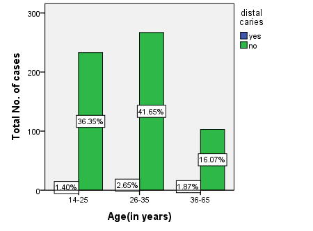
In this study, we observed that there were a total number of 641 subjects out of which 242 subjects were from age 14-25 years (37.8%) and 284 subjects were 26-35 years (44.3%) and 115 subjects were 36-65 years old (17.9%) where the incidence of distal caries were more among the people aged 36-65 years (12/115 patients) [Figure 1].
Figure 1: Bar graph showing association between age distribution of patients with impacted mandibular third molar and presence/absence of distal caries in the adjacent second molar. X-axis: Age of patient (in years) and Y-axis: Total number of cases. Majority of patients of all age groups did not have distal dental caries on the mandibular second molar adjacent to the impacted mandibular molar. However among 115 patients in the age group of 36-65 years, 12 patients had distal dental caries (chi-square test, p-0.043, p<0.05, significant).
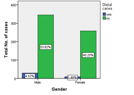
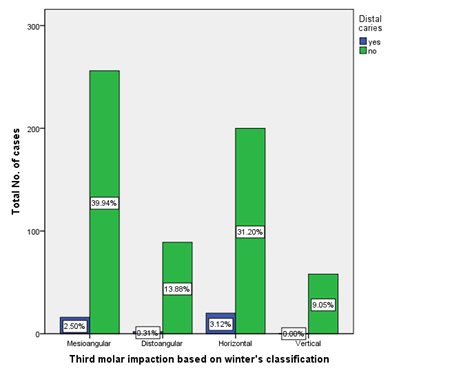
In our study, we can observe that 374 subjects were males and 267 subjects were females. Males (4.52%) had more association to distal caries than females (1.40%) [Figure 2]. Results from our study also showed that horizontal impaction (3.12%) was commonly associated with the presence of distal deep caries followed by mesioangular (2.50%) and distoangular (0.31%). Distal caries were not identified in vertically impacted teeth and results obtained are statistically significant (p<0.05) [Figure 3].
Figure 2:Bar graph showing association between gender distribution of patients with impacted mandibular third molar and presence/absence of distal caries in the adjacent second molar. X-axis: Gender and Y-axis: Total number of cases. Majority of male (53.82%) and female populations (40.25%) did not have distal dental caries on the mandibular second molar adjacent to the impacted mandibular molar (chi-square test, p-0.021, p<0.05, significant). Prevalence of distal dental caries was significantly higher among male population.
Figure 3:Bar graph showing association between the type of mandibular third molar impaction and presence/absence of distal caries on the adjacent second molar. X-axis: Type of mandibular third molar impaction based on winter’s classification; Y-axis: Total number of patients with and without distal caries on adjacent second molar. Totally, 5.9% of the cases were associated with presence of distal dental caries on adjacent second molars. The most common type of impaction associated was horizontal impaction (chi-square value-9.873, p-value-0.020, p<0.05, statistically significant). The association was statistically significant.
Similar study done by Marques et al. showed that distal caries were significantly more frequent when the third molar was in a horizontal position and CEJ was 7-12 mm apart. [25] Another study done by Syed et al. shows mesioangular impaction was the most prominent type and closely followed by horizontal impaction. In their study, age group 21-28 years and males had the higher prevalence of distal caries in the second molar due to impacted third molar. [26]
However in the present study, distal deep caries due impacted teeth was more commonly seen among the elder age groups. The deep distal caries would have occurred due to the presence of the impacted tooth over a longer period of time. With the increase in age their risk for systemic conditions is higher which would invite the need for additional care. Proper diagnosis and treatment planning at an earlier stage will help in prevention of distal caries prior to its occurrence which can preserve the tooth structure and prevent its loss.
Diagnosis of distal caries can be done by using instruments like explorer using tactile sense, by using pulp vitality tests such as electric pulp testing, heat test etc, [27] subjective signs where patient presents with moderate/severe pain in the region of second molar can be seen. Early detection of distal caries can be done by CT scans and periapical images of the teeth. Conebeam computed tomography can render cross-sectional (cut plane) and 3 Dimensional (3D) images that are highly accurate and quantifiable. [28] RMGIC has been found to be the effective material for the restoration in terms of marginal adaptation. [29]
Caries in the second molar could be prevented by prophylactic mandibular third molar extraction that has an angulation of 40°-80° with a contact point on cementoenamel junction to improve the prognosis of mandibular second molars and thus benefit the masticatory function and improve the quality of life. [30] The distal caries can be restored with a biocompatible restorative material depending on the extent of caries. When the caries extends into the pulp, the decision on retaining the tooth depends upon the gingival extent of the caries. If the caries extends below the CEJ, the prognosis is compromised. At times hemisection of the affected second molar is performed to retain the tooth structure and the surrounding alveolar bone. This procedure is done, in case the caries extends only to the distal root of the 2nd molar. This method would facilitate the placement of fixed prosthesis, after extraction of the impacted third molar. [31]
The final line of treatment to prevent complications due to 3rd molar is extraction of the severely affected tooth. Extraction of the impacted third molar has an impact on the adjacent second molar which can compromise the strength of it. Advanced local anesthesia techniques like CCLAD can be used for a painless tooth removal. [32-39] Replacement such as single unit fixed dental prosthesis or implant can be placed to prevent masticatory complications such as the supra eruption of the opposing tooth. Our institution is passionate about high quality evidence based research and has excelled in various fields.
The results of this study can be used as baseline data for future studies involving impacted third molars. Previously our team had conducted numerous clinical trials [40-45] and in vitro studies [46-50] over the past 5 years. Now we are focusing on epidemiological studies. [51-53] The idea for this study stemmed from current interest in our community. Many times, patients do not come with a complaint of impaction, in these cases patients can be informed regarding the possibility of caries in the mandibular second molar due to the impacted mandibular third molar. There is a lack of consideration towards hygiene procedures in patients with impacted teeth, so the patient's motivation and oral hygiene instructions need to be given to the patient to maintain a self-cleansing area and periodic recall, and a follow-up visit to the dentist for caries detection is essential.
Conclusion
Within the limitations of the study, it was concluded that 5.93% of patients with impacted mandibular third molar had distal dental caries on the mandibular second molar. This association was more often identified in the age group of 36-65 years, with relatively higher predilection in the male population. Considering the type of impaction, distal caries on the mandibular second molar was commonly seen in the cases of horizontally impacted third molars.
REFERENCES
- Falci SGM, de Castro CR, Santos RC, de Souza Lima LD, Ramos-Jorge ML, Botelho AM, et al. Association between the presence of a partially erupted mandibular third molar and the existence of caries in the distal of the second molars. Int J Oral Maxillofac Surg. 2012;41(10):1270-4.
- Rajendran R, Kunjusankaran RN, Sandhya R, Anilkumar A, Santhosh R, Patil SR. Comparative evaluation of remineralizing potential of a paste containing bioactive glass and a topical cream containing casein phosphopeptide-amorphous calcium phosphate: An in vitro study. Pesquisa Brasileiraem Odontopediatria e Clinica Integrada. 2019;19(1):1-10.
- Linden W, Cleaton P, Lownie M. Diseases and lesions associated with third molars. Review of 1001 cases. Oral Surg Oral Med Oral Pathol Oral Radiol Endod. 1995;79(2):142-5.
- McArdle LW, Renton TF. Distal cervical caries in the mandibular second molar: An indication for the prophylactic removal of the third molar? Br J Oral Maxillofac Surg. 2006;44(1):42-5.
- Chang SW, Shin SY, Kum KY, Hong J. Correlation study between distal caries in the mandibular second molar and the eruption status of the mandibular third molar in the Korean population. Oral Surg Oral Med Oral Pathol Oral Radiol Endod. 2009;108(6):838-43.
- Kang F, Huang C, Sah MK, Jiang B. Effect of eruption status of the mandibular third molar on distal caries in the adjacent second molar. J Oral Maxillofac Surg. 2016;74(4):684-92.
- Kumar D, Delphine S. Calcified canal and negotiation-A review. Res J Pharma Tech. 2018;11(8):3727.
- Mercier P, Precious D. Risks and benefits of removal of impacted third molars. A critical review of the literature. Int J Oral Maxillofac Surg. 1992;21(1):17-27.
- Charuakkra A, Prapayasatok S, Janhom A, Pongsiriwet S, Verochana K, Mahasantipiya P. Diagnostic performance of cone-beam computed tomography on detection of mechanically-created artificial secondary caries. Imaging Sci Dent. 2011;41(4):143-50.
- Ponnulakshmi R, Shyamaladevi B, Vijayalakshmi P, Selvaraj J. In silico and in vivo analysis to identify the antidiabetic activity of beta sitosterol in adipose tissue of high fat diet and sucrose induced type-2 diabetic experimental rats. Toxicol Mech Methods. 2019;29(4):276-90.
- Mathew MG, Samuel SR, Soni AJ, Roopa KB. Evaluation of adhesion of Streptococcus mutans, plaque accumulation on zirconia and stainless steel crowns, and surrounding gingival inflammation in primary molars: randomized controlled trial. Clin Oral Investig. 2020;24(9):3275-80.
- Subramaniam N, Muthukrishnan A. Oral mucositis and microbial colonization in oral cancer patients undergoing radiotherapy and chemotherapy: A prospective analysis in a tertiary care dental hospital. J Investig Clin Dent. 2019;10(4):e12454.
- Girija ASS, Shankar EM, Larsson M. Could SARS-CoV-2-induced hyperinflammation magnify the severity of Coronavirus disease (Covid-19) leading to acute respiratory distress syndrome? Front Immunol. 2020;11:1206.
- Dinesh S, Kumaran P, Mohanamurugan S, Vijay R, Singaravelu DL, Vinod A, et al. Influence of wood dust fillers on the mechanical, thermal, water absorption and biodegradation characteristics of jute fiber epoxy composites. J Polym Res. 2020;27(1).
- Thanikodi S, Singaravelu D Kumar, Devarajan C, Venkatraman V, Rathinavelu V. Teaching learning optimization and neural network for the effective prediction of heat transfer rates in tube heat exchangers. Therm Sci. 2020;24(1B):575-81.
- Murugan MA, Jayaseelan V, Jayabalakrishnan D, Maridurai T, Kumar SS, Ramesh G, et al. Low velocity impact and mechanical behaviour of shot blasted SiC wire-mesh and silane-treated aloevera/hemp/flax-reinforced SiC whisker modified epoxy resin composites. Silicon Chem. 2020;12(8):1847-56.
- Vadivel JK, Govindarajan M, Somasundaram E, Muthukrishnan A. Mast cell expression in oral lichen planus: A systematic review. J Investig Clin Dent. 2019;10(4):e12457.
- Chen F, Tang Y, Sun Y, Veeraraghavan VP, Mohan SK, Cui C. 6-shogaol, an active constiuents of ginger prevents UVB radiation mediated inflammation and oxidative stress through modulating NrF2 signaling in human epidermal keratinocytes (HaCaT cells). J Photochem Photobiol B. 2019;197:111518.
- Manickam A, Devarasan E, Manogaran G, Priyan MK, Varatharajan R, Hsu C-H, et al. Score level based latent fingerprint enhancement and matching using SIFT feature. Multimed Tools Appl. 2019;78(3):3065-85.
- Wu F, Zhu J, Li G, Wang J, Veeraraghavan VP, Krishna Mohan S, et al. Biologically synthesized green gold nanoparticles from induce growth-inhibitory effect on melanoma cells (B16). Artif Cells Nanomed Biotechnol. 2019;47(1):3297-305.
- Ma Y, Karunakaran T, Veeraraghavan VP, Mohan SK, Li S. Sesame inhibits cell proliferation and induces apoptosis through inhibition of STAT-3 translocation in thyroid cancer cell lines (FTC-133). Biotechnol Bioprocess Eng. 2019;24(4):646-52.
- Ponnanikajamideen M, Rajeshkumar S, Vanaja M, Annadurai G. In vivo type 2 diabetes and wound-healing effects of antioxidant gold nanoparticles synthesized using the insulin plant Chamaecostus cuspidatus in albino rats. Can J Diabetes. 2019;43(2):82-9.e6.
- Vairavel M, Devaraj E, Shanmugam R. An eco-friendly synthesis of Enterococcus sp.-mediated gold nanoparticle induces cytotoxicity in human colorectal cancer cells. Environ Sci Pollut Res Int. 2020;27(8):8166-75.
- Paramasivam A, VijayashreePriyadharsini J, Raghunandhakumar S. N6-adenosine methylation (m6A): A promising new molecular target in hypertension and cardiovascular diseases. Hypertens Res. 2020;43(2):153-4.
- Marques J, Montserrat-Bosch M, Figueiredo R, Vilchez-Perez M-A, Valmaseda-Castellon E, Gay-Escoda C. Impacted lower third molars and distal caries in the mandibular second molar. Is prophylactic removal of lower third molars justified? J Clin Exp Dent. 2017;9(6):e794-8.
- Syed KB, Alshahrani FS, Alabsi WS, Alqahtani ZA, Hameed MS, Mustafa AB, et al. Prevalence of distal caries in mandibular second molar due to impacted third molar. J Clin Diagn Res. 2017;11(3):ZC28-30.
- Janani K, Palanivelu A, Sandhya R. Diagnostic accuracy of dental pulse oximeter with customized sensor holder, thermal test and electric pulp test for the evaluation of pulp vitality: An in vivo study. Braz Dent Sci. 2020;23(1):8.
- Ramanathan S, Solete P. Cone-beam computed tomography evaluation of root canal preparation using various rotary instruments: An in vitro study. J Contemp Dent Pract. 2015;16(11):869-72.
- Nasim I, Hussainy S, Thomas T, Ranjan M. Clinical performance of resin-modified glass ionomer cement, flowable composite and polyacid-modified resin composite in noncarious cervical lesions: One-year follow-up. J Conserv Dent. 2018;21(5):510-515.
- Prajapati VK, Mitra R, Vinayak KM. Pattern of mandibular third molar impaction and its association to caries in mandibular second molar: A clinical variant. Dent Res J (Isfahan). 2017;14(2):137-142.
- Alqahtani SM. Tooth hemisection: Case study and literature review. Int J Med Dent. 2019;23(2).
- Baskran RNR, Pradeep S. Recent advancement of local anasthesia advancement to recent advancement of local anaesthesia administration. J Pharm Res. 2016;
- Priyadharsini VJ. In silico validation of the non-antibiotic drugs acetaminophen and ibuprofen as antibacterial agents against red complex pathogens. J Periodontol. 2019;90(12):1441-8.
- Ezhilarasan D, Apoorva VS, Ashok Vardhan N. Syzygium cumini extract induced reactive oxygen species-mediated apoptosis in human oral squamous carcinoma cells. J Oral Pathol Med. 2019;48(2):115-21.
- Ramesh A, Varghese S, Jayakumar ND, Malaiappan S. Comparative estimation of sulfiredoxin levels between chronic periodontitis and healthy patients-A case-control study. J Periodontol. 2018;89(10):1241-8.
- Mathew MG, Samuel SR, Soni AJ, Roopa KB. Evaluation of adhesion of Streptococcus mutans, plaque accumulation on zirconia and stainless steel crowns, and surrounding gingival inflammation in primary molars: Randomized controlled trial Clin Oral Investig. 2020;24(9):3275-3280.
- Sridharan G, Ramani P, Patankar S, Vijayaraghavan R. Evaluation of salivary metabolomics in oral leukoplakia and oral squamous cell carcinoma. J Oral Pathol Med. 2019;48(4):299-306.
- Pc J, Marimuthu T, Devadoss P. Prevalence and measurement of anterior loop of the mandibular canal using CBCT: A cross sectional study. Clin Implant Dent Relat Res. 2018;20(4):531-534.
- Ramadurai N, Gurunathan D, Samuel AV, Subramanian E, Rodrigues SJL. Effectiveness of 2% Articaine as an anesthetic agent in children: randomized controlled trial. Clin Oral Investig. 2019;23(9):3543-50.
- Nallaswamy D, Solete P, Subha M. Comparative study on conventional lecture classes versus flipped class in teaching conservative dentistry and endodontics. Int J Res Pharma Sci. 2019;10(1):689-93.
- Jose J, Subbaiyan H. Different treatment modalities followed by dental practitioners for ellis class 2 fracture-A questionnaire-based Survey. Open Dent J. 2020;14:59-65.
- Janani K, Ajitha P, Sandhya R. Improved quality of life in patients with dentin hypersensitivity. Saudi Endo J. 2020;10:81.
- Noor S, Others. Chlorhexidine: Its properties and effects. Res J Pharma Tech. 2016;9(10):1755-60.
- Hussainy SN, Nasim I, Thomas T, Ranjan M. Clinical performance of resin-modified glass ionomer cement, flowable composite and polyacid-modified resin composite in noncarious cervical lesions: One-year follow-up. J Conserv Dent. 2018;21(5):510-5.
- Teja KV, Ramesh S, Priya V. Regulation of matrix metalloproteinase-3 gene expression in inflammation: A molecular study. J Conserv Dent. 2018;21(6):592-6.
- Ravinthar K. Recent advancements in laminates and veneers in dentistry. Res J PharmaTechn. 2018;11(2):785-7.
- Rajakeerthi R, Ms N. Natural Product as the storage medium for an avulsed tooth-A Systematic review. Cumhuriyet Dental Journal. 2019;22(2):249-56.
- Pradeep AR, Shemesh H, Jothilatha S, Vijayabharathi R, Jayalakshmi S, Kishen A. Diagnosis of vertical root fractures in restored endodontically treated teeth: A time-dependent retrospective cohort study. J Endod. 2016;42(8):1175-80.
- Ramamoorthi S, Nivedhitha MS, Divyanand MJ. Comparative evaluation of postoperative pain after using endodontic needle and endo activator during root canal irrigation: A randomized controlled trial. AustEndod J. 2015;41(2):78-87.
- Teja KV, Ramesh S. Shape optimal and clean more. Saudi Endo J. 2019;9(3):235.
- Siddique R, Sureshbabu NM, Somasundaram J, Jacob B, Selvam D. Qualitative and quantitative analysis of precipitate formation following interaction of chlorhexidine with sodium hypochlorite, neem and tulsi. J Conserv Dent. 2019;22(1):40-7.
- Manohar M, Sharma S. A survey of the knowledge, attitude, and awareness about the principal choice of intracanal medicaments among the general dental practitioners and nonendodontic specialists. Indian J Dent Res. 2018;29(6):716-720.
- Nandakumar M, Nasim I. Comparative evaluation of grape seed and cranberry extracts in preventing enamel erosion: An optical emission spectrometric analysis. J Conserv Dent. 2018;21(5):516-20.


 The Annals of Medical and Health Sciences Research is a monthly multidisciplinary medical journal.