Cleidocranial Dysplasia with Autosomal Dominant Inheritance Pattern
- *Corresponding Author:
- Dr. Saba Khan
Department of Oral Medicine and Radiology, NIMS Dental College and Hospital, NIMS University, Shobha Nagar, Jaipur-Delhi Highway, Jaipur - 303 121, Rajasthan, India.
E-mail: dr.sabakhan23@gmail.com
Abstract
Cleidocranial dysplasia (CCD) is an autosomal dominant disease with a wide range of expression, characterized by clavicular hypoplasia, retarded cranial ossification, delayed bone and teeth development, supernumerary teeth, stomatognathic, craniofacial and skeletal abnormalities. This paper presents a case of CCD in a female with brachycephalic skull, depressed frontal bone and nasal bridge, hypoplastic middle one‑third of face with mandibular prognathism and hyper mobility of both shoulders with associated radiographic features. Odontologist is often the first professional who patient of CCD approaches, since there is a delay in the eruption or absence of permanent teeth. The premature diagnosis allows a scope for proper treatment modalities, offering a better life quality for patient.
Keywords
Clavicle, Cleidocranial dysplasia, Genetic disorder, Supernumerary teeth
Introduction
Cleidocranial Dysplasia (CCD) is a rare congenital disorder of autosomal dominant inheritance with prevalence of 0.5/100,000 live births.[1,2] CCD primarily affects bones that undergo intramembranous ossification. It is also known as Marie and Sainton disease, Scheuthauer-Marie-Sainton syndrome, mutational dysostosis and cleidocranial dysostosis.[3] These patients have the following triad of lesions, which is considered pathognomonic for its diagnosis such as multiple supernumerary teeth, partial or complete absence of the clavicles and open sagittal sutures and fontanelles.[4]
Case Report
A 26-year-old female reported to dental out-patient department with the chief complaint of mobility of upper front teeth since two months. Patient was healthy and mentally alert.
Extra oral examination revealed frontal bossing with high supercilliary arches and increased interpupillary distance.
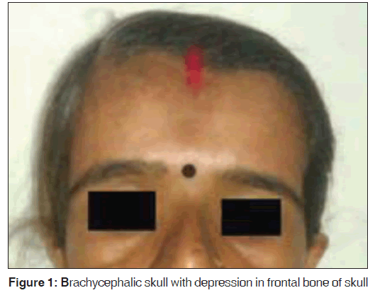
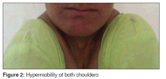
Brachycephalic skull with depression in the frontal bone of skull, depressed nasal bridge and hypoplastic middle one-third of face with mandibular prognathism was evident [Figure 1]. Physical examination of the chest revealed hypoplastic clavicle on both sides with hyper mobility of both shoulders and ability to move them toward midline [Figure 2]. She gave the history that among the other family members her father, elder brother and elder daughter were having similar physical attributes.
On intra-oral examination, there were no soft-tissue abnormalities. The palate was narrow and high arched. There was grade III mobility in respect to 12. Many missing teeth were evident in both maxillary and mandibular arch. Based on the clinical findings a provisional diagnosis of CCD, localized periodontitis with 12 was considered.
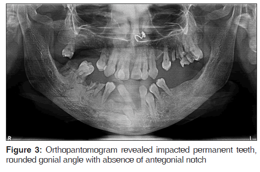
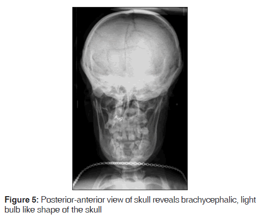
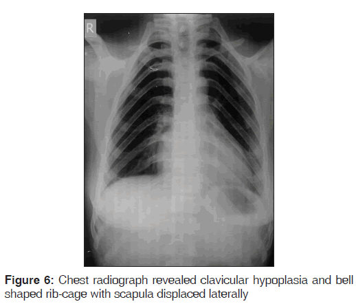
Orthopantomogram was advised, which revealed numerous impacted permanent teeth. Other finding includes rounded gonial angle with the absence of antegonial notch. Sigmoid notch was deepened with lack of proper outline of condylar and coronoid process. There was near parallelism of anterior and posterior border of ramus of mandible [Figure 3]. Lateral cephalogram revealed non-closure of sutures due delayed ossification of anterior fontanel, small wormian bones and enlargement of occiput was also obvious. Hypoplastic maxilla, depressed nasal bridge, non-pneumatization of frontal sinuses [Figure 4]. Posterior anterior view of skull reveals widened cranium and open anterior and posterior fontanelles. Brachycephalic skull results in the light bulb like shape to the silhouette of the skull [Figure 5]. Chest radiograph confirmed the clavicular hypoplasia and bell shaped rib-cage with scapula displaced laterally [Figure 6].
Considering the above mentioned clinical and radiographic findings a final diagnosis of CCD was made. Patient 12 was extracted and she was recalled for further follow-ups and her Figure 1: Brachycephalic skull with depression in frontal bone of skull Figure 3: Orthopantomogram revealed impacted permanent teeth, rounded gonial angle with absence of antegonial notch Figure 5: Posterior-anterior view of skull reveals brachycephalic, light bulb like shape of the skull daughter was scheduled for a follow biannual follow-up to review the status of her erupting permanent or impacted teeth.
Discussion
In 1898, Mane and Sainton were the first to describe cases of this disease and associated them with patterns of inheritance. Later Bauer and Kallialla suggested genetic mutation as an etiological factor with an autosomal dominant inheritance, as seen in the present case where the patients’ father and daughter both showed similar physical characteristics. However, in some cases external interferences in the fetal period could cause this mutation that is transferred to the progeny.[3]
The CCD gene has been mapped to chromosome 6p21 within a region containing the core binding factor-α1 (CBFA1) gene. CBFA1 controls differentiation of precursor cells into osteoblasts and is thus essential for membranous as well as endochondral bone formation, which may be related to delayed ossification of skull, teeth, pelvis and extremities.[5]
CCD presents with skeletal defects of several bones, the most striking of which are partial or complete absence of clavicles, late closure of fontanels, presence of open skull sutures and multiple wormian bones. The skull base is dysplastic and the growth is reduced, which results in increased skull width leading to brachycephaly and hypertelorism.[6] Delayed closure of anterior fontanel and metopic sutures result in frontal bossing. Bossing of the frontal, occipital and parietal regions give the skull a large globular shape with a small face. The characteristic skull abnormalities are sometimes referred to as the “Arnold head.”
Other facial features include broad and depressed nasal bridge, narrow high arched palate, hypertelorism and absent paranasal sinuses due to underdeveloped maxilla. The above enlisted features were observed in our patient also. The thoracic cage is small with short ribs, with underdeveloped clavicles to varying degrees and in approximately 10% of cases, clavicles are completely absent. Chest may be narrowed or funnel-shaped leading to potential respiratory distress in childhood. Other bones are also affected including long bones, vertebral column, pelvis and bones of hand and feet.[7] Decreased eruptive force causes prolonged retention of deciduous teeth and impaction of permanent teeth. The failure in the eruption can be related with the absence of cellular cementum in the roots of permanent teeth or lack of absorption of the deciduous teeth or presence of fibrous connective tissue interposed between the dental follicle and the mucosa.[8]
The radiological features of CCD are also very characteristic. The cranial abnormalities include wide-open sutures, patent fontanels, presence of wormian bones and delayed ossification of skull, decreased pneumatization of paranasal, frontal and mastoid sinuses, impacted supernumerary teeth. Chest X-ray shows cone shaped thorax with narrow upper thoracic diameter, absent or hypoplastic clavicles and scapulae.[9]
Pseudoarthrosis can be considered as differential diagnosis, but it is evidenced by hypoplasia or absence of one of the clavicle predominantly right side clavicle. CCD can be differentiated from Pyknodysostosis as the latter shows features such as malformed bones, osteosclerosis, delayed suture closures, anodontia, delayed eruption of permanent teeth and dysplasia of distal phalanges.[4] Gardner’s syndrome, which has multiple intestinal polyposis, multiple sebaceous cysts and multiple osteomas distinguishes Gardner’s syndrome from CCD.
Conclusion
CCD patients often present with impacted teeth, which may have a propensity to develop dentigerous cysts in future, so it is always advisable for the dental professional to keep a scheduled follow-up for the screening of these patients. The proposed treatment options are restoration and preservation of the deciduous teeth and orthognathic surgery to fix the maxillary hypoplasia. Implant placements is another therapeutic option in the oral rehabilitation of these patients.[10]
Source of Support
Self-funded.
Conflict of Interest
None declared.
References
- Yochum TR, Rowe LJ. Essentials of Skeletal Radiology, 2nd ed. Baltimore: Williams and Wilkins; 1996. p. 589-92.
- Brueton LA, Reeve A, Ellis R, Husband P, Thompson EM, Kingston HM. Apparent cleidocranial dysplasia associated with abnormalities of 8q22 in three individuals. Am J Med Genet 1992;43:612-8.
- Kalliala E, Taskinen PJ. Cleidocranial dysostosis. Report of six typical cases and one atypical case. Oral Surg Oral Med Oral Pathol 1962;15:808-22.
- Tanaka JL, Ono E, Filho EM, Castilho JC, Moraes LC, Moraes ME. Cleidocranial dysplasia: Importance of radiographic images in diagnosis of the condition. J Oral Sci 2006;48:161-6.
- Shankar YU, Reddy R, Swetha D. Cleidocranial dysplasia: A case report. Int J Contemp Dent 2011;2:73-5.
- Golan I, Baumert U, Hrala BP, Müssig D. Dentomaxillofacial variability of cleidocranial dysplasia: Clinicoradiological presentation and systematic review. Dentomaxillofac Radiol 2003;32:347-54.
- Goaz PW, White SC. Oral Radiology: Principles and Interpretation. Elsevier publications, 5th ed, 2004.
- Rushton MA. An anomaly of cementum in cleidocranial dysostosis. Br Dent J 1956;100:81.
- Feldman VB. Cleidocranial dysplasia: A case report. J Can Chiropr Assoc 2002;46:185-91.
- Dawjee SM, Nkhumeleni F. Cleidocranial dysplasia – A case report. SADJ 2002;57:187-90.




 The Annals of Medical and Health Sciences Research is a monthly multidisciplinary medical journal.