Comparison of Glycogen Positive Cells in Oral Smears with Random Blood Sugar Levels of Type 2 Diabetes Patients
2 Department of Oral Pathology and Microbiology, Vishnu Dental College, Bhimavaram, West Godavari, Andhra Pradesh, India
Citation: Reddy M, et al. Comparison of Glycogen Positive Cells in Oral Smears with Random Blood Sugar Levels of Type 2 Diabetes Patients. Ann Med Health Sci Res. 2018; 8: 1-5
This open-access article is distributed under the terms of the Creative Commons Attribution Non-Commercial License (CC BY-NC) (http://creativecommons.org/licenses/by-nc/4.0/), which permits reuse, distribution and reproduction of the article, provided that the original work is properly cited and the reuse is restricted to noncommercial purposes. For commercial reuse, contact reprints@pulsus.com
Abstract
Background: Even though much advancement in the medical field, diagnosis of diabetes mellitus (DM) needs an invasive procedure for drawing of blood and estimation of blood sugar levels. The purpose of this study was to analyse the glycogen content in exfoliated cells of oral mucosa as an adjunct in the diagnosis of diabetes mellitus. Aim: To assess the number of PAS (Periodic Acid Schiff) positive glycogen containing cells in oral smears and correlating the findings with random blood sugar levels. Subjects and Methods: Smears were taken from buccal mucosa of 60 type II diabetes mellitus patient and 60 healthy individuals. The smears were stained with Periodic Acid Schiff stain. Each smear was screened for 100 random cells and number of PAS positive cells was noted. Based on the staining intensity of the cells, PAS positive cells are categorized as intense, moderateand mild PAS positive. Numbers of PAS positive cells were correlated with serum glucose levels of each patient. Statistical analysis used is Student T test and Pearson correlation. Results: Student t test and Pearson correlation was conducted which reveals Intense and moderate PAS positive cells was significantly higher (p<0.001) in the study group and also has significant correlation with random blood sugar levels and total PAS positive cells. Conclusion: Correlating the obtained results suggests that random blood glucose levels are one of the factors responsible for the increased glycogen positive cells from oral smears. Demonstration of increased PAS positive cells can be used as a non-invasive diagnostic screening tool for DM provided the study is repeated with larger sample of subjects.
Keywords
Type II diabetes; Periodic acid Schiff staining; Oral cytological smears; Random blood sugar levels
Introduction
Diabetes mellitus is fast growing and a potential epidemic in India with more than 62 million diabetic individuals currently diagnosed with the disease. [1] According to Wild et al. the prevalence of diabetes is predicted to double globally with 171 million in 2000 to 366 million in 2030 with a maximum increase in India. [2]
Diabetes mellitus (DM) is one of the major endocrine metabolic disorders characterized by hyperglycemia due to relative or absolute insulin deficiency resulting in decreased utilization of glucose by body cells. [3] Oral effects of diabetes include xerostomia, dental caries, epithelial atrophy, ulcerations, delayed wound healing, infections (candidiasis), burning mouth syndrome (BMS) and periodontal destruction. [3-5] Early screening in high risk group and frequent monitoring of glycemic levels in known diabetics will reduce the complication occurring due to the diabetes mellitus. [6,7]
Frequent monitoring the blood glucose levels in a diabetic patient involves invasive procedure to draw the blood such as venipuncture and finger lancing which is painful. Due to above reasons a simple non-invasive or minimal invasive monitoring are gaining interest in present situation. One such technique is exfoliative cytology which is simple, rapid, inexpensive, bloodless and painless procedure in which physiologically exfoliated cells from the mucous membrane are used as efficient diagnostic adjunct in varied oral lesions with better patient acceptability. [8-11] The purpose of this study is to demonstrate glycogen in exfoliated cells of buccal mucosa in type II diabetics and correlate the obtained results with serum glucose levels to establish exfoliative cytology as a diagnostic adjunct in diabetes mellitus. We also have assessed the additional finding of fungal positivity in the oral smears stained with PAS and also their relation with serum glucose levels.
Subjects and Methods
This is a case control study which was conducted in the Department of Oral Pathology and Microbiology, Vishnu dental college, Bhimavaram, Andhrapradesh, India. Total of 120 subjects were taken into consideration of which Study group consisting of 60 (33 females and 27 males) known type II Diabetic patients with the disease duration ranging from 1 year to 12 years. Control group consist of 60 healthy, age and sex matched subjects. Patients with habits, oral lesions and other systemic disorders were excluded from the study. Patients were explained about the study, informed consent was obtained and detailed case histories with duration of disease, medication were recorded.
2 ml of patients’ venous blood was drawn under aseptic condition for estimation of random blood glucose levels using GOD-POD enzymatic method, spectrophotometer (biozyme liquid glucose kit, Robonik, Mumbai).
Examination of oral cavity was done; patients and controls were asked to rinse the mouth thoroughly with water. Scrapings were taken from buccal mucosa using a disposable wooden spatula by exerting gentle pressure. The scraps were smeared on to the clean, fresh and non-greasy slide in a circular pattern over a large area preventing clumping of cells. The slides were immediately fixed using liquid spray fixative (Biofix). All the slides were stained with Periodic Acid Schiff (PAS) technique.
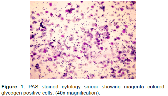
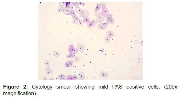
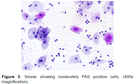
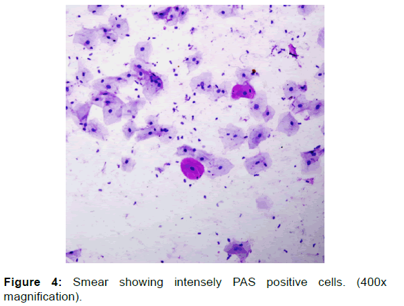
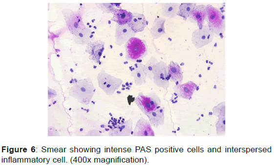
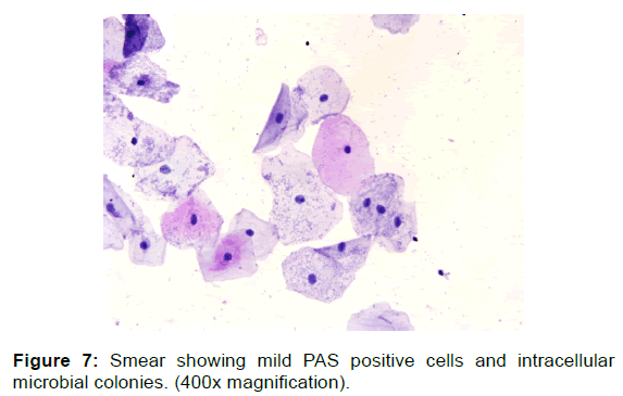
PAS stained slides were screened in a zigzag manner and each slide was analysed for magenta colored glycogen positive cells [Figure 1] using Olympus BX 51 research microscope and image pro plus software. Approximate 100 cells were viewed at magnification of 100x and the values were given as number of PAS positive cells/100 cells. Each cell counted was analysed for the staining intensity and the cells were named as mild PAS positive [Figure 2], moderate PAS positive [Figure 3] and intensely PAS positive cells [Figure 4].
The obtained variables were then compared with serum glucose levels of each patient. Student t test and Pearson Correlation was used to assess the statistical significance among the variables of the test and control groups.
Results
The study group included 45% males and 55% females with an age range of 35-65 years. Study group consisted of patients with history of diabetes ranging from 1 to 12 years. Fifty percent of patients had the disease duration for less than 5 years. All the patients were under oral hypoglycemic. RBS levels in the study group ranged from 124 to 245 mg/dl, with an average of 186.60 ± 60.71 mg/dl.
The comparison of RBS levels, total PAS positive cells, intense PAS, moderate PAS and mild PAS positive cells of study and control groups along with p values were represented in Table 1.
| Variables | Study group | Control group | P-value |
|---|---|---|---|
| RBS value | 179.07 ± 53.856 | 112.80 ± 23.35 | <0.001** |
| Total PAS cells | 32.53 ± 10.649 | 15.30 ± 7.02 | <0.001** |
| Total Intense positive cells | 10.08 ± 6.442 | 4.00 ± 3.67 | 0.001** |
| Total medium positive cells | 9.28 ± 4.830 | 6.05 ± 3.56 | 0.021* |
| Total low positive cells | 13.17 ± 6.82 | 5.25 ± 3.83 | <0.001** |
Table 1: The comparison of RBS levels, total PAS positive cells, intense PAS, moderate PAS and mild PAS positive cells of study and control groups along with p values.
Student t test done for the variables compared between study and control groups were strongly significant with p value of less than or equal to 0.001 except for the total moderate PAS positive cells with p value of 0.021 which was also significant.
Pearson correlation test done for RBS values and intensity of PAS positive cells within the study group revealed significant correlation between RBS and total PAS positive cells with P< 0.04. No correlation was found between RBS and intense, medium and mild PAS positive cells [Table 2].
| Variables | Correlation coefficient | Probability |
|---|---|---|
| RBS and total PAS cells | 0.057 | 0.04 |
| RBS and total intense positive cells | 1.98 | 0.130 |
| RBS and total medium positive cells | 1.53 | 0.24 |
| RBS and total mild positive cells | 0.25 | 0.11 |
Table 2: Pearson correlation between RBS and intense, medium and mild PAS positive cells in study group.
Discussion
Diabetes mellitus is the most common endocrine disorder distinguished by increase in blood glucose levels due to relative or absolute insulin deficiency which needs routine monitoring of blood glucose for effective management and prevention of complications. Monitoring of blood glucose levels is an invasive method, which is a costly and time consuming. Exfoliate cytology is non-invasive technique having good patient compliance, cost effective and an effective method for screening large populations. [8-11]
Glycogen, the primary storage form for glucose in mammalian cells acts as energy reserve, the formation of which is regulated by insulin, adrenaline and glucagon hormones. These hormones fail to break down glycogen, leading to accumulation of glycogen in the cells and tissues. [12,13]
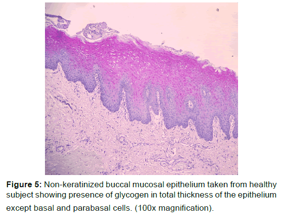
Varying amount of glycogen is found in upper layers of normal buccal mucosa especially in prickle, granular and surface cells excluding basal and Para basal layers [Figure 5]. The glycogen rich cells were most pronounced in the regions over the rete ridges [14,15] but the glycogen in the epithelial cells decreases in hyperkeratosis and leukoplakic mucosa. [16] Histochemical study of diabetic gingiva revealed more widely distributed intensely reactive PASpositive material when compared to non-diabetic controls. [17]
The present study analyzed the significance of the glycogen positive cells in the exfoliated buccal mucosal cells as an adjunctive diagnostic aid in diabetic patients. Here total PAS positive cells were counted in all the smears of study, control groups revealed significant increase in number of glycogen positive cells in study group (p=0.001) which was similar to the study result by Hallikerimath et al., [18] Asemi-Rad et al., [19] Yasminsatpathy et al., [20] Raty Ravindran et al., [21] and Latti et al. [22] When intensity of PAS positive cells and number of PAS positive cell were compared there was significant increase in diabetics than controls, this was similar to the study conducted by Asemi-Rad et al., [19] RatyRavindran et al. [21]
PAS positive cell staining intensity in our study was subdivided into intense, moderate, mild PAS positive cells. Current study demonstrated higher number of mild and intensely stained cells than moderately PAS positive cells in diabetic and control group. Pearson Correlation was done between random blood sugar levels and intensity of staining i.e., total PAS positive cells, intensely, moderately and mild PAS positive cells. Found significant correlation (p=0.04) between random blood sugar levels and total PAS positive cells, rest of variable showed insignificant correlation, this is in contrast to the study done by RatyRavindran et al. [21]
Poor serum glucose control in diabetics makes the patients oral cavity prone to infections. [23,24] We observed increased amount of inflammatory cell infiltrate [Figure 6] and increased intracellular microbial colonies [Figure 7] in the diabetics than the control groups.
Oral candidiasis is the most common and prevalent among diabetics than in non-diabetics. [25] Among 60 PAS stained smears of diabetics considered for the study only 3 smears revealed candida hyphae of which 1 smear showed high amount hyphae. When random blood sugar levels and candidal hyphae were compared, we found increased amount of hyphae [Figure 8] in patient with increased RBS levels.
Conclusion
Exfoliative cytology is one of the non-invasive and patient friendly procedures which have good patient compliance, rapid, cost effective method for screening of large group of population. Routine screening of blood glucose in diabetic patients is necessary to maintain and prevent the complications occurring secondary to the diabetes. From the results of study conducted reveal that exfoliative cytology in conjunction with PAS staining can be used as an adjunctive diagnostic aid for routine chair side screening which is a noninvasive technique which can bypasses the multiple venipuncture’s and pain associated with it for the diabetic patient. However, the study with larger samples should be conducted to come to a definitive conclusion.
Acknowledgements
We all thank our teachers and well-wishers Dr. T.R. Saraswathi, Dr. C.R. Ramachandran for their continuous guidance and support throughout the study period
Conflict of Interest
All authors disclose that there was no conflict of interest.
REFERENCES
- Kaveeshwar SA, Cornwall J. The current state of diabetes mellitus in India. Australasian Medical Journal 2014; 7: 45-48.
- Wild S, Roglic G, Green A, Sicree R, King H. Global prevalence of diabetes: estimates for the year 2000 and projections for 2030. Diabetes Care 2004; 27:1047-1053.
- Al-Maskari AY, Al-Maskari MY, Al-Sudairy S. Oral manifestations and complications of diabetes mellitus: A review. SQU Medical Journal 2011; 11: 179-186.
- Little JW, Falace DA, Miller CS, Rhodis NL. Dental management of the medically compromised patient. 6th ed. St. Louis: Mosby; 2002; 248-268.
- Greenburg MS, Glick M. Burket’s oral medicine: Diagnosis and treatment. 10th ed. Hamilton: BC Decker; 2003; 563‑572.
- Mohan V, Sandeep S. Epidemiology of type 2 diabetics: Indian scenario. Indian J Med Res 2007; 125: 217-230.
- Ramachandran A, Snehalatha C, Viswanathan V. Burden of Type 2 diabetes and its complications—the Indian scenario. Curr Sci 2002; 83: 1471-1475.
- Ziskin DE, Siegel EH, Loughlin WC. Diabetes in relation to certain oral and systemic problems – clinical study of dental caries, tooth eruption, gingival changes, growth phenomena, and related observations in juveniles. J Dent Res 1942; 21: 296.
- Nayar AK, Sivapathasundharam B. Cytomorphometric analysis of exfoliated normal buccal mucosa cells. Indian J Dent Res 2003; 14: 87-93.
- Rickles NH. Oral exfoliative cytology: An adjunct to biopsy. CA Cancer J Clin 1972; 22:163-171
- Alberti S, Cesar TS. Exfoliative cytology of the oral mucosa in type II diabetic patients: Morphology and cytomorphometry. JOPM 2003; 32: 538-545
- Kishi K. Cytochemical study of exfoliated cells of oral mucosa. Acta Med Okayama 1975; 29: 103-109.
- Bouche C, Shanti Serdy C, Kahn R, Allison B. Goldfine. The cellular fate of glucose and its relevance in Type 2 diabetes. Endocrine Reviews 2004; 25: 807-830
- Merwyn A, Hubert L, Schroeder E. Differentiation in normal human buccal mucosa epithelium. J Anat 1979; 128: 31-51.
- Xiao T, Kurita H, Li X, Qi F, Shimane T, Aizawa H, et al. Iodine penetration and glycogen distribution in vital staining of oral mucosa with iodine solution. Oral Surg Oral Med Oral Pathol Oral Radiol 2014; 117: 754-759.
- Silverman S, Barbosa J, Kearns G. Ultrastructural and histochemical localization of glycogen in human normal and hyperkeratotic oral epithelium. Archs oral Biol 1971; 16: 423-434.
- Kronman JH, Cohen MM, Colte D, Waitzken L. Histologic and histochemical study of human diabetic gingiva. J Dent Res 1970; 49: 177.
- Hallikerimath S, Sapra G, Kale A, Malur PR. Cytomorphometric analysis and assessment of periodic acid Schiff positivity of exfoliated cells from apparently normal buccal mucosa of type 2 diabetic patients. Acta Cytol 2011; 55: 197-202.
- Asemi-Rad A, Heidari Z, Mahmoudzadeh-Sagheb H. The relationship between the staining intensity of oral exfoliative cells with periodic acid schiff and cytomorphometric indices with fasting blood sugar in type 2 Diabetic patients. Interdisciplinary J Contemp Research Business 2013; 4: 375-381.
- Satpathy Y, Kumar PS. Promising role of exfoliative cytology in the evaluation of glyceamic status of typr II diabetics: a pilotstudy. J. Maxillofac Oral Surg 2015; 14: 206-211.
- Ravindran R, Gopinathan DM, Sukumaran S. Estimation of salivary glucose and glycogen content in exfoliated buccal mucosal cells of patients with type II diabetes mellitus. Journal of Clinical and Diagnostic Research 2015; 9: 89-92.
- Latti BR, Birajdar SB, Latti RG. Periodic acid Schiff-Diastase as a key in exfoliative cytology in diabetics: a pilot study. Journal of Oral and Maxillofacial Pathology 2015; 19: 188-191.
- Trott JR. The presence and distribution of glycogen in the gingiva. Aust Dent J 1957; 2: 283-294
- Li X, Kolltveit KM, Tronstad L, Olsen I. Systemic diseases caused by oral infections. Clin Microbiol Rev 2000; 13: 547-558
- Pallavan B, Ramesh V, Dhanasekaran BP, Sudipindu N, Govindarajan V. Comparison and correlation of candida colonization in diabetic patients and normal individuals. Journal of Diabetes & Metabolic Disorders 2014; 13: 66.



 The Annals of Medical and Health Sciences Research is a monthly multidisciplinary medical journal.