Endomyocardial Fibrosis in Northern Nigerian Girl
- *Corresponding Author:
- Dr. Ibrahim Aliyu
Department of Pediatrics, Aminu Kano Teaching Hospital, Bayero University, Kano, Nigeria.
E-mail: ibrahimaliyu2006@yahoo. com
Abstract
Endomyocardial fibrosis (EMF) was first described as far back as 1946 among Ugandans. It is mostly a disease of the tropics and subtropics and most common among individuals of low socio‑economic class affecting an estimated 10 million people.[1] In Nigeria it has been reported among individuals in the southern part, in the rain forest zone.
https://marmaris.tours
https://getmarmaristour.com
https://dailytourmarmaris.com
https://marmaristourguide.com
https://marmaris.live
https://marmaris.world
https://marmaris.yachts
Keywords
Endomyocardial fibrosis, Hausa girl, Northern Nigeria
Introduction
Endomyocardial fibrosis (EMF) was first described as far back as 1946 among Ugandans. It is mostly a disease of the tropics and subtropics and most common among individuals of low socio-economic class affecting an estimated 10 million people.[1] In Nigeria it has been reported among individuals in the southern part, in the rain forest zone. Though very few cases have been documented in Guinea and Sudan savannah.[2] It has bimodal peak age distribution of 10 and 30 years. However its incidence and demographic characteristics is not completely known in northern Nigeria because of its believed rarity in the savannah region. I therefore report a case of EMF in a 10-year-old northern Nigerian girl of a middle class family who had never left the shores of Kano-Jigawa axis.
Case Report
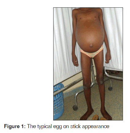
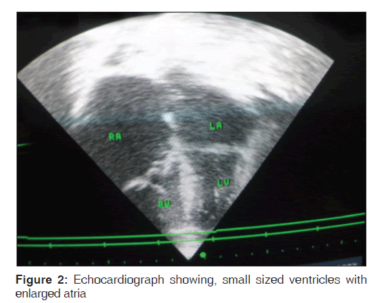
A 10-year-old girl first seen at the age of 8-years with the complaint of abdominal swelling with occasional shortness of breath for which she had been seen severally in a general hospital. She was the third child of the parents five children in a monogamous family setting, both parents were health workers and there was no similar complaint in the family; she had Bacillus Calmette and Guerin BCG vaccine at birth and was even treated for abdominal tuberculosis during the course of the illness. On examination, she was wasted with a weight of 20 kg (61% of expected for age), she was not cyanosed and she had no per-orbital or pedal edema and no digital clubbing. The pertinent findings in the cardiovascular system were; tachycardia with a pulse rate of 120 b/min, the blood pressure was normal (100/70 mmHg) sitting right arm, the jugular venous pressure was elevated (9 cmH2O), the precordium was normo-active with the apex-beat poorly appreciated and distant heart sounds, she also had pansystolic murmurs maximal at the left sternal margin grade 4/6 and at the apical area radiating toward the axilla grade 4/6 with bilateral posterior basal crepitations; the abdominal examination revealed massive ascites [Figure 1]. Full blood count was not remarkable. Chest X-ray showed a globular heart; electrocardiogram showed small QRS complex in V1. An ALOKA prosound SSD 4000 ultrasound system, with facilities for M-mode, 2D, color flow mapping and Doppler studies was used for echocardiography; all measurements were done using 5.0 MHz sector transducer. Echocardiograph [Figure 2] showed enlarged left atrium with a diameter of 37 mm (24-35 mm). The mitral valves were thickened with tethering of the supporting apparatus with mitral regurgitation; the E: A ratio of the mitral valve inflow was three with deceleration time of 120 ms. The ejection fraction was 65% and fractional shortening of 34%. The left ventricle was of normal size with left ventricular diameter in end-diastole and systole of 36 mm and 23 mm respectively (34-43 mm and 21-29 mm respectively). There was obliterated of the right ventricular apex however no thrombus was seen; the right ventricular diameters in end-diastole and systole were 32 mm and 22 mm respectively with fractional area change of 38% (32-60%); E: A ratio of 2.5 and deceleration time of 110 ms. The tricuspid valves were also regurgitant and thickened with tethering of the valvular apparatus, but the outflow tracts were spared. The right atrium was enlarged with a minor axis of 46 mm (29-45 mm). The liver and renal function tests were not remarkable.
Discussion
EMF is the most common cause of restrictive cardiomyopathy among Africans, but its exact cause still eludes researchers despite first described over 60-years ago. Though there is a dearth of study in Northern Nigeria in recent times, several hypothesis postulated in the past were later discarded for lack of evidence; such as the impact of plantain consumption-serotonin hypothesis; the effect of cerium-geophysical hypothesis; but the helminthes-eosinophilia hypothesis has received wider acceptance. There is often great similarity in the cardiac pathology in patients with Loffler’s carditis and EMF, therefore much emphasis is placed on the role of eosinophilia in its etiopathogenesis.
EMF may affect the right, left or both ventricles. Our case had both ventricular involvements. There is often poor association between the clinical and structural abnormalities in most cases and some patients may remain asymptomatic until late into the illness.[3] Late presentation of diseases is often a problem in resource limited countries therefore most of the early features which could help in disease definition are often missed; such as the presence of eosinophilia therefore absence of eosinophilia was not surprising in this case. It is generally believed that EMF is rare in the northern part of Nigeria however this case adds up to two cases reported within a span of 2 years in our institution[2] despite a reported decreasing incidence in the southern part of Nigeria.[4] Therefore clinician should be alert to the possibility of a changing trend in the epidemiology of this disease.
This case was quite unique because she developed the disease at an early age of the 8-years, more so she was from a middle income family, which made malnutrition as a possible contributing factor unlikely; this further compound the controversies surrounding its etiopathogenesis, therefore this highlights the need for extensive studies to determine the exact cause of the disease.
EMF can easily be confused with other common causes of ascites and weight loss in children such as chronic liver disease and tuberculosis especially where the tuberculosis score is routinely applied, as was the case in this patient; therefore, it should be considered as a differential of ascites not only in adolescents and young adults, but also in school age children irrespective of the place of domicile in Nigeria.
EMF has no specific treatment and it carries a poor prognosis with mean survival of 2 years; this index case had been on digoxin, furosemide and had remained stable for 2 years so far..
Conclusion
EMF is a common cause of restrictive cardiomyopathy among Africans seen in the rain forest and rarely seen in the Sahel or Guinea savannah. However we may be seeing a change in the trend in the disease pattern, which calls for a community based study on the prevalence of the disease in the savannah region; furthermore, it should be considered as a possible differential of ascites in school age children.
Source of Support
Nil.
Conflict of Interest
None declared.
References
- Yacoub S, Kotit S, Mocumbi AO, Yacoub MH. Neglected diseases in cardiology: A call for urgent action. Nat Clin Pract Cardiovasc Med 2008;5:176-7.
- Mijinyawa MS, Sani MU. Endomyocardial fibrosis presenting outside the tropical rain forest. J Natl Med Assoc 2010;102:1258-60.
- Salemi VM, Rochitte CE, Barbosa MM, Mady C. Images in cardiology. Clinical and echocardiographic dissociation in a patient with right ventricular endomyocardial fibrosis. Heart 2005;91:1399.
- Akinwusi PO, Odeyemi AO. The changing pattern of endomyocardial fibrosis in South-west Nigeria. Clin Med Insights Cardiol 2012;6:163-8.


 The Annals of Medical and Health Sciences Research is a monthly multidisciplinary medical journal.