Epithelial Inclusion Cyst in a Cervical Lymph Node: Report of a Rare Entity at an Uncommon Location
- *Corresponding Author:
- Jetley S
Department of Pathology, Hamdard Institute of Medical Sciences and Research, Jamia Hamdard, New Delhi, India
E-mail: drjetley2013@gmail.com
This is an open access article distributed under the terms of the Creative Commons Attribution-NonCommercial-ShareAlike 3.0 License, which allows others to remix, tweak, and build upon the work non-commercially, as long as the author is credited and the new creations are licensed under the identical terms.
Dear sir,
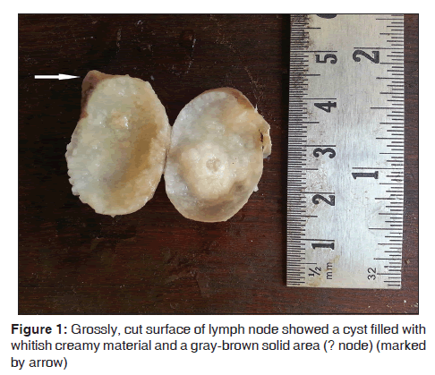
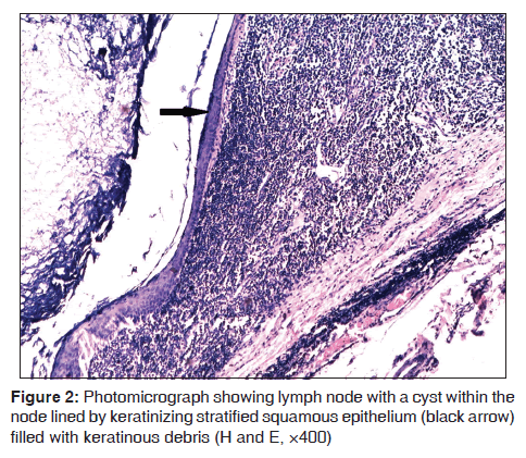
Benign inclusions in lymph nodes are foci of nonneoplastic ectopic tissue, which may be of various types. The most worrisome feature of these benign inclusions in lymph node is that they may be mistakenly interpreted as tumor metastasis.[1,2] Therefore, the awareness of such an entity is all the more important in preventing overdiagnosis of a malignant lesion. We hereby present a case, clinically suspected to be tuberculosis, which surprisingly turned out to be a benign epithelial inclusion cyst within a submandibular lymph node.A 16-year-old female presented with right submandibular swelling without associated pain, fever, or respiratory symptoms. There was no past or family history of tuberculosis. A single, nontender, 2.5 cm × 2.0 cm right submandibular lymph node was palpable. A clinical diagnosis of tubercular lymphadenopathy was considered. Hematological and biochemical investigations and chest X-ray were normal. Erythrocyte sedimentation rate was 22 mm in 1st h. Mantoux test revealed an induration of 8 mm. The patient had a fine needle aspiration cytology (FNAC) report (4 months old) suggestive of reactive lymphadenitis from a private laboratory. She subsequently refused a repeat FNAC. The lymph node was then excised and a single, capsulated lymph node measuring 2.5 cm × 2.0 cm × 1 cm was received. Cut surface showed a cyst filled with whitish creamy material. At the periphery on one of the poles, there was a gray-brown solid area (? node) [Figure 1]. Microscopic sections showed the capsule of the lymph node with underlying compressed lymphoid follicles along with a cyst within the lymph node lined by keratinizing stratified squamous epithelium filled with keratinous debris [Figure 2]. There was no evidence of atypia, loss of polarity, or increased mitosis within the lining of the cyst. A diagnosis of an epithelial inclusion cyst within a cervical lymph node was rendered.
Benign inclusions in lymph nodes were first described by Ries in 1897.[2] Brooks et al.[3] classified them into three different types: Epithelial, nevomelanocytic, and decidual. Epithelial cells in these inclusions may originate from breast tissue, salivary gland tissue, squamous epithelial cells, thyroid tissue, etc. Fellegara et al.[4] classified nodal epithelial inclusions into three categories: Glandular type (most common), squamous type inclusions (least common), and mixed glandular-squamous type inclusions.
The most commonly involved lymph nodes vary with the type of heterotopic inclusions. Breast tissue and nevus cells are more commonly encountered in axillary nodes whereas salivary gland tissue and thyroid tissue are usually found in cervical nodes. Decidual tissue, intestinal glands, or mesothelial cells are seen in pelvic and retroperitoneal nodes. The origin of epithelial inclusions is debatable. The various theories proposed are embryogenic, implantation (iatrogenic), and metaplastic origins.[1,2]
Detailed morphological evaluation of lymph nodes is crucial in reaching at an accurate diagnosis as these inclusions may be misinterpreted as tumor metastasis. The criteria favoring benign inclusions include bland nuclear cytology, absence of anaplasia, brisk mitosis, and necrosis.
On extensive search of the literature, we came across only a few reports of squamous inclusion cysts in cervical nodes.[5,6] We considered it worthwhile to report this rare occurrence in a submandibular lymph node to create awareness among young pathologists about the existence of such an unusual benign entity in a lymph node. Awareness of this rare entity can prevent misdiagnosis of a malignant lesion and thereby unnecessary overzealous treatment.
Financial support and sponsorship
Nil.
Conflicts of interest
There are no conflicts of interest.
References
- Ioachim HL, Medeiros LJ, editors. Epithelial inclusions in lymph nodes. In: Ioachim’s Lymph Node Pathology. 4th ed. Philadelphia: Lippincott Williams & Wilkins; 2009. p. 283-8.
- Spinardi JR, Goncalves IR, La Falce TS, Fregnani JH, Barros MD, Macea JR. Benign inclusions in lymph nodes. Int J Morphol 2007;25:625-9.
- Brooks JS, LiVolsi VA, Pietra GG. Mesothelial cell inclusions in mediastinal lymph nodes mimicking metastatic carcinoma. Am J Clin Pathol 1990;93:741-8.
- Fellegara G, Carcangiu ML, Rosai J. Benign epithelial inclusions in axillary lymph nodes: Report of 18 cases and review of the literature. Am J Surg Pathol 2011;35:1123-33.
- Fruehwald-Pallamar J, Li CQ, Hasteh F, Hauff S, Davidson TM, Mafee MM. Nodal inclusion cyst in a cervical lymph node. Neurographics 2012;2:163-6.
- Tomasino RM, Perino A. Epithelial inclusions in the lymph nodes. Study of 3 cases with submandibular localization. Arch De Vecchi Anat Patol 1975;60:529-45.


 The Annals of Medical and Health Sciences Research is a monthly multidisciplinary medical journal.