Evaluation of Effect of Etching Duration and Loading Conditions on Fracture Resistance of Ceramic Veneers Prepared on Permanent Maxillary Central Incisors
Published: 27-Aug-2021
Citation: Apte A and Kambala SS*. D Evaluation of Effect of Etching Duration and Loading Conditions on Fracture Resistance of Ceramic Veneers Prepared on Permanent Maxillary Central Incisors-An Experimental Study. AMHSR. 2021;11:118-121
This open-access article is distributed under the terms of the Creative Commons Attribution Non-Commercial License (CC BY-NC) (http://creativecommons.org/licenses/by-nc/4.0/), which permits reuse, distribution and reproduction of the article, provided that the original work is properly cited and the reuse is restricted to noncommercial purposes. For commercial reuse, contact reprints@pulsus.com
Abstract
Background: Using ceramic veneers for restoration of non-esthetic teeth has magnified with the increased demand of patient awareness. Several factors affect prolonged prognosis of ceramic veneers. This study will help to comparatively evaluate the effect of etching duration and loading conditions on the resistance to fracture of ceramic veneers. Objectives: To evaluate the fracture resistance of “Lithium disilicate ceramic veneers” at an angle of 60⁰ after exposure to 5% hydrofluoric acid for 20 secs and 40 secs then comparative evaluation of the same. Methodology: Study comprises of 12 samples of freshly extracted permanent maxillary central incisors followed by mounting them in self polymerizing acrylic resin, and preparing butt joint design tooth preparation. Tooth preparation followed by making its impression using polyvinyl siloxane impression material with a customized tray and poured with die stone. Wax pattern will be fabricated over it for fabrication of veneers. After fabrication, etchant application will be done for a specific period of 20 sec & 40 sec over the prepared tooth followed by applying bonding agent. Cementation procedure will be followed. The mounted teeth will be exposed to a load at an angulation of 60⁰ simulating intercostal movement and will be evaluated and compared for resistance to fracture by using Universal Testing Machine. Expected results: This study will help the dentists to assess better fracture resistance of ceramic veneers. Conclusion: With the help of this study dentists can use effectiveness of optimum etching duration on ceramic veneers.
Keywords
Ceramic veneers; Incisal butt joint; Etching duration
Introduction
Until about the last two decades in dental practice, esthetics now is equally necessary as function, biology and structure as current advertising and media in general enhance pleasant appearance because of its importance in the day to day activities.
One of the major aspects of dental treatment is to imitate the teeth and design smiles in a most natural and esthetic way based on particular needs of any individual.
Anterior region restorations are viewed to be a complex step in cosmetic dentistry where the appropriate materials are to be selected along with an ideal treatment to get a satisfactory and predictable outcome.
Research of most materials since 1960s is in a way of creating stronger and reinforced restorations with accurate margins however in 1980s there came a new wave of ceramic products.
Dental porcelain has excellent esthetics in combination with biocompatibility and is one of the most commonly used restorative materials. [1]
Over last few years’ new glass ceramic restorative materials have been developed with magnified strength properties along with prudent optical properties making them ideal for fabrication of esthetic veneers and crown. Ceramic veneers are the routine conservative esthetic treatment of abnormally formed, malaligned, discolored; traumatized, fractured and worn anterior teeth are selected to give exemplary esthetics. The structure of veneering ceramic has been stated as an amorphous and glass matrix comprises a random network of cross-linked silica in a tetrahedral arrangement which is inbuilt in different amounts of non-dissolved feldspar and reinforcement crystals like lithium disilicate. [2] All-ceramic restorations gained popularity because of its improved physical and esthetic properties and clinical success. [3]
Several factors govern the ceramic restoration’s accomplishment clinically like preparation design, constituents of the material and its cementing procedure. Combination of etching to be done with hydrofluoric acid succeeded by silane coupling agent achieves ideal bonding to “lithium disilicate glass ceramic”. [4] So, by considering this aspect of ceramics, present study is planned for evaluation of effect of etching duration with loading conditions on fracture resistance of ceramic veneers.
Restoration of anterior teeth that are non-esthetic has always faced a difficulty in practice for dental practitioner. The use of ceramic laminate veneers for restoration of teeth that are non-esthetic has magnified with the growing demand of awareness of patient. There are several factors affecting prolonged prognosis of the ceramic veneers like selection of case, surface of tooth, design prepared, thickness of material, laboratory veneers fabrication, cementation material and functional and para functional situations. [5] After viewing literature various studies have been conducted with the above mentioned aspects, but till now no study has been conducted to see the result of comparative evaluation of the mentioned features.
Ceramic veneers have become a popular treatment modality due to their conservative and esthetic nature and clinical performance. [6] All-ceramic systems have expanded their range of indications in almost all the areas of fixed restorative dentistry. [7] So this present study will help to comparatively evaluate the effect of etching duration and loading conditions on resistance to fracture of ceramic veneers.
Aim
To evaluate the effect of the etching duration and loading condition on fracture resistance of ceramic veneers prepared on permanent maxillary central incisors
Objective
• To evaluate the fracture resistance of Lithium disilicate ceramic veneers at an angle of 60⁰ after exposure to 5% hydrofluoric acid for 20 sec.
• To evaluate the fracture resistance of Lithium disilicate ceramic veneers at an angle of 60⁰ after exposure to 5% hydrofluoric acid for 40 sec.
• Comparative evaluation of the fracture resistance of Lithium disilicate ceramic veneers at an angle of 60⁰ after exposure to 5% hydrofluoric acid for 20 sec and 40 sec.
Material and Methods
This is an experimental type of study.
Duration-2 years
Sample size-6 PER GROUP
Statistical analysis-
Statistical analysis will be done by using response analysis Student’s unpaired t- test, Student’s paired t-test and Chi square test. The software using the analysis will be SPSS 24.0 version and p< 0.05 is considered as the level of significance
Formula-
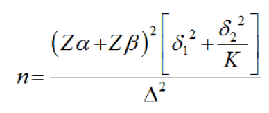
Sample size formula for difference between two means:

Where,
Zα is the level of significance at 5% i.e. 95%
Confidence interval=1.96
Zβ =Power of the test=80%=0.84%
δ1=SD of fracture load values in subgroup [1A=39.08]
δ2 = SD of fracture load values in subgroup [1B=41.80]

=5.49
=6 patients needed in each group
Formula Reference:
VK Chadha, Sample Size Determination in Health Studies,
NTI Bulletin, 2006,42/3 and 4,55-62. [8] [Figure 1]
Inclusion criteria
• Freshly extracted permanent maxillary central incisors with proper morphology of the tooth.
• The teeth should be caries free.
Exclusion criteria
Restored, fractured, deciduous teeth and teeth with fluorosis.
Data collection tools-Universal testing machine with which fracture resistance of ceramic veneers can be calculated.
Materials required
• E-Max Lithium dislocate ingots
• Etchant
• Bonding agent
• Polyvinyl siloxane impression material
• Light body impression material
• Phosphate bonded investment material
• Self-polymerizing acrylic resin
Equipment required
• Custom made metallic mold
• Surveyor
• Universal testing machine
• Custom made impression tray
Methodology
This is an in vitro study, which will be conducted in Department of Prosthodontics, Sharad Pawar Dental College, Sawangi (Meghe), Wardha.
It will be comprising of 12 samples of freshly extracted permanent maxillary central incisors. A customized stainless steel mould in two-piece will be used for fabricating resin blocks of standard size in which mounting of the specimen will be done.
Proper positioning of the specimen and mould will be done with the help of a surveyor followed by mixing of self-cure acrylic resin powder with its liquid and ultimately pouring it around the tooth specimen till the mould is filled completely.
Mounting of the extracted permanent maxillary central incisors in self polymerizing acrylic resin will be done, followed by butt joint design tooth preparation for partial veneers. The tooth will be prepared and its impression will be made using polyvinyl siloxane impression material with a customized tray and poured with die stone and wax pattern will be made over it for fabrication of veneers with E-max lithium disilicate material.
Samples will be divided in two groups:
Group A-Etching to be done with Hydrofluoric acid for 20 sec
Group B-Etching to be done with Hydrofluoric acid for 40 sec
After fabrication of the veneers, application of etchant will be done for a specific pre decided durations that is 20 sec and 40 sec over the prepared extracted tooth followed by applying bonding agent and cementation procedure. Ultimately the mounted teeth will be exposed to a load at an angulation of 60⁰ simulating intercuspal movement and will be comparatively evaluated for etching duration and loading conditions on fracture resistance by using universal testing machine.
Expected Outcomes/Results
The expected outcome is that the clinicians will have a better idea about the time of exposure that should be carried out in order to achieve better fracture resistance of ceramic veneers at the loading condition of 60⁰.
Discussion
Lucas Villaca Zogheib et al. carried out a study where they examined different effectiveness of etching time on roughness and flexural strength of glass ceramic that is lithium disilicate based. Specimens were taken as ceramic bars (16mm x 2mm x 2mm) produced from blocks of ceramic. Polishing of all specimens was done and cleaned sonically with distilled water. They divided the specimens into five groups randomly in which A Group was control group, B Group to E gone under surface treatment with 4.9% hydrofluoric acid for various etching durations: 20s, 60s, 90s, 180s respectively. Etched surfaces were observed with the help of scanned electron microscopy. They concluded that the increased HF acid etching time showed an effect on rough surface and the flexural strength of “lithium disilicate based glass ceramic”. [2]
Burcin Akogul et al in 2010 carried out a study on 75 central incisors which were extracted and divided according to five preparation designs as 2mm reduction of incisal surface completely in the enamel, 4mm reduction of incisal surface completely in the enamel, 2mm reduction of incisal surface completely in the dentin, 4mm reduction of incisal surface completely in the dentin and teeth that are not restored, control as intact teeth and came to a conclusion that preparation of ceramic veneers completely in dentin with 4mm reduction of incisal surface gave less fracture loads than with 2 mm reduction of incisal surface. [3]
Rontani et al in 2017 carried out a study in which they evaluated the effect of various concentration of hydrofluoric acid in association with different etching durations onto resin cement’s strength to bond to a “lithium disilicate glass ceramic” and concluded that minimum hydrofluoric acid concentration of 5% and proper bond with lithium disilicate ceramic is made in time of 20secs. [4]
Aman Arora et al in 2017 conducted a study in which they evaluated the effectiveness of the incisal butt joint and the incisal overlap design under loading conditions that is 125⁰ and 60⁰ on the resistance to fracture of ceramic veneers, 125⁰ simulating protrusive and 60⁰ simulating intercuspal movements. They came to a conclusion that palatal overlap showed lesser fracture resistance than butt joint design and was higher at 125⁰ than 60⁰. [5]
Sy Yin Chai et al in 2019 carried out a study for evaluation of load to failure of different design preparations that is “butt joint” preparation and “feathered edge” preparation of ceramic veneers and correlated these outcomes to failure mode of restorations. Load to failure values of ceramic veneers showed important outcomes by loading angulations and incisal preparation designs. Feathered edge group exhibited lesser load to failure value as compared to butt joint group. [6] Few other related studies were reviewed. [9,10]
Limitations: The study is restricted only to freshly extract permanent maxillary central incisors that are anterior teeth which are not restored, fractured and deciduous. The design preparation used in the study is butt joint only. Acrylic resin that will be used will not be able to simulate the intraoral conditions.
Conclusion
With the help of this study dental practitioners can use effectiveness of etching duration and loading conditions when ceramic veneers are to be made. This will help to satisfy the patients’ needs according to the esthetics and time span of the ceramic veneer in the oral cavity.
References
- Belkhode VM, Nimonkar SV, Godbole SR, Nimonkar P, Sathe S, Borle A. Evaluation of the effect of different surface treatments on the bond strength of non-precious alloy-ceramic interface: An SEM study. J Dent Res Dent Clin Dent Prospects. 2019;13:1-8.
- Zogheib LV, Bona AD, Kimpara ET, Mccabe JF. Effect of hydrofluoric acid etching duration on the roughness and flexural strength of a lithium disilicate-based glass ceramic. Braz Dent J. 2011;22:45-50.
- Akoğlu B, Gemalmaz D. Fracture resistance of ceramic veneers with different preparation designs. J Prosthodont. 2011;20:380-384.
- Puppin-Rontani J, Sundfeld D, Costa AR, Correr AB, Puppin- Rontani RM, Borges GA.et,al Effect of hydrofluoric acid concentration and etching time on bond strength to lithium disilicate glass ceramic. Oper Dent. 2017;42:1-10.
- Arora A, Upadhyaya V, Arora SJ, Jain P, Yadav A. Evaluation of fracture resistance of ceramic veneers with different preparation designs and loading conditions: An in vitro study. J Indian Prosthodont Soc. 2017;17:1-7.
- Chai SY, Bennani V, Aarts JM, Lyons K, Lowe B. Effect of incisal preparation design on load‐to‐failure of ceramic veneers. J Esthet Restor Dent. 2020;32:424-432.
- Dudhekar AU, Nimonkar SV, Belkhode VM, Borle A, Bhola R. Enhancing the esthetics with all-ceramic prosthesis. J Datta Meghe Inst Med Sci U. 2018;1:13155.
- Chadha VK. Sample size determination in health studies. NTI bulletin. 2006;42:2-8.
- Vyas R, Suchitra SR, Gaikwad PT, Gurumurthy V, Arora S, Shetty S. Assessment of fracture resistance capacity of different core materials with porcelain fused to metal crown: An in vitro study. J Contemp Dent Pract. 2018;19:389-392.
- Sancheti Y, Kambala S, Godbole S, Kambala R, Dhamande M, Pisulkar S. Effect of multiple firing on flexural strength and color stability of pressable all ceramic material: An invitro study. J Datta Meghe Inst Med Sci U. 2020;15:94-97.


 The Annals of Medical and Health Sciences Research is a monthly multidisciplinary medical journal.