Giant Cell Fibroma of Tongue: A Case Report
Received: 07-Mar-2023, Manuscript No. AMHSR-23-97581; Editor assigned: 10-Mar-2023, Pre QC No. AMHSR-23-97581(PQ); Reviewed: 24-Mar-2023 QC No. AMHSR-23-97581; Revised: 27-Apr-2023, Manuscript No. AMHSR-23-97581(R); Published: 25-May-2023
Citation: Zarbade RV, et al. Giant Cell Fibroma of Tongue: A Case Report. Ann Med Health Sci Res. 2023;13:680-682.
This open-access article is distributed under the terms of the Creative Commons Attribution Non-Commercial License (CC BY-NC) (http://creativecommons.org/licenses/by-nc/4.0/), which permits reuse, distribution and reproduction of the article, provided that the original work is properly cited and the reuse is restricted to noncommercial purposes. For commercial reuse, contact reprints@pulsus.com
Abstract
Giant cell fibroma is a rare type of fibroma which appears in the oral cavity and has a distinct histopathology. It accounts for about 2% to 5% of all oral fibrous lesions. Due to the identical clinical features, it can be diagnosed as irritational fibroma, but histological characteristics help to distinguish giant cell fibroma. We present a case of giant cell fibroma of the tongue in a 30- year-old female, which was diagnosed as giant cell fibroma using histological features.
Keywords
Giant cell fibroma; Tongue; Laser; Oral cavity
Introduction
Giant Cell Fibroma (GCF) was first identified as a novel form of fibrous hyperplastic soft tissue in 1974 by Weathers and Calliham. GCF is rarely fibrous lesion found in oral cavity. The large mononuclear and multinucleated giant cells that characterize the tumor have led to the choice of the term giant-cell fibroma. These enormous cells frequently resemble stellate structures and have oval nuclei with eosinophilic cytoplasm.
Weathers and Callihan state that all lesions are less than 1 cm in diameter and, in most cases, less than 0.5 cm. They are often pedunculated and have a nodular or cerebriform surface. In reality, an examination of these fibrous lesions revealed that many of them were clinically diagnosed as papillomas, with the majority described as “fibroepithelial polyp.” None of them were described as pigmented or vascular in nature indicating that they are mucosal in colour.The gingiva was the most prevalent site for the GCF, followed by the buccal mucosa and tongue.Other sites included the lip and the floor of the mouth. The age range of people with GCF varied greatly.The GCF showed no significant sex preference. The fibromas, on the other hand, were more common in females than in males.The GCF is a pedunculated or sessile in nature nodule. It is mostly made up of fibrous connective tissue that is normally loosely distributed, rarely myxomatous, and at times mature and compact.
Vascularity is common, with well-formed capillaries and venules. There is no the epithelium hypertrophy. Unless the epithelium surface is traumatically ulcerated, inflammation is uncommon. When inflammation is present, it is frequently combined, with both acute and chronic cells involved. Under the epithelium, there are many spindle-shaped cells. Sometimes there is an artifactual gap or detachment of the collagen fibres from the cell borders. The cytoplasm is basophilic and granular, with a few vacuoles.
A single nucleus is normally present, however two are not uncommon. The nucleus is big, circular to oval, and fluidfilled, with a fine, evenly distributed granular chromatin structure and a large solitary nucleolus. Some cells can have up to seven or eight nuclei, generating multinucleated giant cells. GCF is most common in Caucasians in their third decades of life, with a slight female predilection. The cause of GCF is unknown [1,2].
Case Presentation
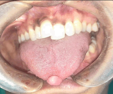
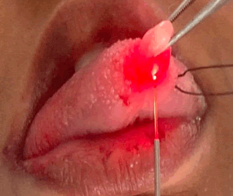
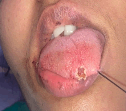
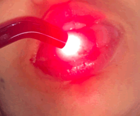
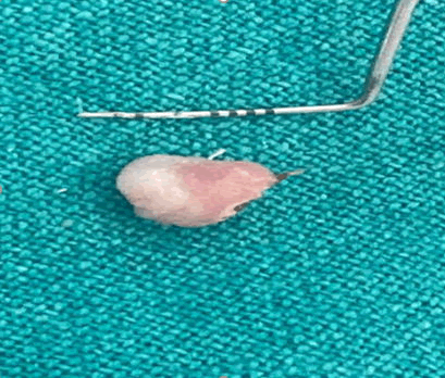
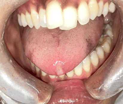
A 30-year-old female patient was reported to the department of periodontology with a complaint of an overgrowth in the tip of her tongue that was growing but not causing pain. The growth was circular in shape, approximately 4 mm 10 mm in diameter, smooth surfaced, usual mucosal colour and pedunculated (Figure 1). It was unyielding and consistent, with no history of trauma. The preliminary diagnosis was irritational fibroma. A local anesthetic injection was given at the base of the mass and was resected using diode laser (Novolase). The procedure was preceeded and followed by photobiomodulation for better healing. Parameters were 980 nm wavelength, 2.5 watt at continuous mode and for photobiomodulation 660 nm 0.2 watt for 2 minutes (Figures 2-5) [3,4].
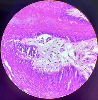
On excisional biopsy, histological specimen revealed the stratified squamous parakeratinized epithelium showing many papillomatous areas. The underlying connective tissue was moderately cellular and showing numerous giant stellate shaped fibroblast. Few endothelial lined blood vessels were also noted (Figure 6). Based on these characteristics, the final diagnosis of giant cell fibroma was made. After a week, the post-operative visit was deemed satisfactory, with no pain and normal tissue healing (Figure 7) [5].
Discussion
The GCF is a rare pathological entity that primarily affects young children, with a higher prevalence among teenagers and young adults in their second and third decades of life. GCF of the oral cavity is a pedunculated fibrous lesion that can range in size from 0.5 cm to 4 cm. GCF is most oftenly found in the oral cavity on gingiva, followed by the tongue and buccal mucosa. The exact cause of GCF is unidentified. The most widely accepted theory currently is that GCF evolved in response to trauma or recurrent chronic inflammation. Because of the lack of a fibrous capsule and a single vascularity, GCF has not been considered a true neoplastic lesion [6,7].
Despite the presence of granulation tissue, there is vascular distension and engorgement, ruling out the possibility that GCF is formed by pyogenic granuloma cells. The presence of inflammatory cells indicates that fibroblastic differentiation was caused by recurrent inflammation. When Weathers and Callihan (1974) reported the first case of GCF, they speculated that the giant cell could be a melanocyte or a Langerhans cell. However, several studies concluded that GCF is of endothelial and myofibroblastic origin, as evidenced by negative staining for alpha-smooth muscle actin. Elastin is found in fibroepithelial polyps but not in GCF, according to immunological and histological studies. This is a significant distinction between fibroepithelial polyps and GCF [8,9].
GCF is treated by excision of the lesion with a scalpel, electro-cautery or laser, but in adults, surgical excision is preferred, whereas in children, electro surgery or laser excision is recommended. When it comes to electrosurgery, there has been a lot of debate. The greatest issue with this technique has been the lateral heat produced within adjacent electro surgery electrodes. Laser therapy is an alternative method for removing soft tissue lesions. Nd:YAG, CO2, and diode lasers are recommended for fibroma removal. The benefits of laser include immediate haemostasis and disinfection of the surgical site, least postoperative pain and inflammation, the elimination of sutures, and the acceleration of the healing process. Recurrence is uncommon, but some cases have been reported. Follow-up is required on a regular basis.
Conclusion
The cells of this fibrous tumour are of two types. One is spindle or stellate type of cell and the other one being multinucleated giant cells. Both these cell types are fibroblastic in origin. This lesion, which usually appear on the gingiva, is more common in younger people. All GCFs can be managed conservatively with no recurrence.
References
- Weathers DR, Callihan MD. Giant-cell fibroma. Oral Surg Oral Med Oral Pathol. 1974;37:374-384.
[Crossref] [Google Scholar] [PubMed]
- Gnepp DR. Diagnostic surgical pathology of the head and neck e-book. 2nd edition, Elsevier Health Sciences, China, 2009.
- Minatel Braga M, Carvalho AL, Vasconcelos MC, Henrique Braz-Silva P, Luiz Pinheiro S. Giant cell fibroma: A case report. J Clin Pediatr Dent. 2006;30:261-264.
[Crossref] [Google Scholar] [PubMed]
- Reibel J. Oral fibrous hyperplasias containing stellate and multinucleated cells. Euro J Oral Sci. 1982;90:217-226.
[Crossref] [Google Scholar] [PubMed]
- Campos E, Gomez RS. Immunocytochemical study of giant cell fibroma. Braz Dental J. 1999;10:89-92.
[Google Scholar] [PubMed]
- Sabarinath B, Sivaramakrishnan M, Sivapathasundharam B. Giant cell fibroma: A clinicopathological study. J Oral Maxillofac Pathol. 2012;16:359.
[Crossref] [Google Scholar] [PubMed]
- Butchi Babu K, Nag S, Hussain MW, Mishra A. Laser excision of giant cell fibroma-A report of a case and review of literature. Ann Essences Dent. 2010;2:221-224.
- Kalkwarf KL, Krejci RF, Edison AR, Reinhardt RA. Lateral heat production secondary to electrosurgical incisions. Oral Surg Oral Med Oral Pathol. 1983;55:344-348.
[Crossref] [Google Scholar] [PubMed]
- Kotlow LA. Lasers in pediatric dentistry. Dent Clin. 2004;48:889-922.
[Crossref] [Google Scholar] [PubMed]



 The Annals of Medical and Health Sciences Research is a monthly multidisciplinary medical journal.