Histopathological Profile of Breast Cancer in an African Population
- *Corresponding Author:
- Dr. Gerald Dafe Forae
Department of Pathology, University of Benin Teaching Hospital, P.M.B. 1111, Benin-City, Edo, Nigeria.
E-mail: jforae2000@yahoo.com
Abstract
Background: Currently breast cancer (BRCA) still remain the most commonly diagnosed female cancer globally with a significant geographic, racial and ethnical variations in its incidence. Aim: This article examines the frequency and histological types and grades of BRCA in a pioneer teaching Hospital in Delta State, Nigeria. Materials and Methods: H and E stained‑slides of breast biopsies diagnosed at the Central Hospital, Warri from 2005 to 2011 were archived and studied. Request forms were scrutinized for clinical bio‑data, diagnosis and histological sections were analyzed. Data obtained were analyzed using the Statistical Package for the Social Sciences version 17 statistical package (SPSS) Incorporated, Chicago, Illinois, USA, and value presented descriptively. Results: During this period, 905 breast lesions were seen. Out of this, 261 were BRCAs, of which 260 cases were females and one case was a male. The peak age incidence for BRCA and its variants was 40‑49 years accounting for (n = 94/261; 36%). The mean age of BRCA was 46 years (6.2). Invasive carcinoma of no special type (NST) was the most commonly encountered histological group of breast carcinoma constituting (n = 203/261, 77.7%) with the high grade invasive ductal carcinoma as the leading diagnosis. Conclusion: Majority of BRCAs encounter was invasive ductal carcinoma of NST with bulk of patients presenting in Stages III and IV.
Keywords
Histopathology, Invasive ductal carcinoma, Pre-menopausal, Staging
Introduction
Breast cancer (BRCA) is a disease of antiquity. About 1550 B C. BRCA was first documented in the Ebers papyrus of the ancient Egyptians as cited by S.M. Arab in medicine in Ancient Egypt.[1] Until date, BRCA still remain the most commonly diagnosed female cancer accounting for 20% of female malignancies globally.[2,3] This does not mean that BRCA cannot occur in males and children as quite a number of cases have been reported world-wide.[4] Globally, there are significant geographic, racial and ethnical variations in its incidence.[5] Reports have it that BRCA incidence and mortality rate is higher in developed communities when compared with developing communities.[6] Presently in the United States alone, it was estimated that one in every eight and one in every 14 Caucasian-American and African-Americans female respectively will develop BRCA in her lifetime. Again the United States have an average whopping 207,090 incidence cases of BRCA yearly.[6] Studies have it that Asians and Africans have the lowest incidence rate globally.[2,3,6] In Japan and other far Asian countries, the incidence rate of BRCA is five times less than their American counterparts.[7] However, in spite of this, BRCA still remain top among other female cancers in both continents.[3,6,7] BRCA incidence and mortality rates in West African countries is more common when compared with East African countries.[2,6] In Nigeria, the exact national incidence rate is not known because of lack of comprehensive data, however, it ranked among the first two most common female cancers.
This study intends to describe the frequency, stages, histological patterns, staging and grading of BRCA in a local scenario of a tertiary Hospital Institution in Southern Nigeria as it compares with other parts of Nigeria and elsewhere.
Materials and Methods
The Central Hospital is a referral Hospital located in Warri. It is one of the most patronized Government Hospitals in South-South Nigeria, providing health care services for more than three million people in Delta State and its environs. It also served as the pioneer teaching hospital to the Delta State University with the Pathology Department fully accredited for histopathological services. This study is a descriptive study using retrospective review of 905 breast biopsies records of surgical pathology daybooks of the Pathology Departments from 2005 to 2011. Ethical approval was obtained from the Warri Central Hospital ethical committee.
All specimens sent for histology include (biopsies, mastectomies and wide excision biopsies with lymph nodes) were fixed in 10% formalin solution, processed with histokinette automated tissue processor, paraffin embedded and sectioned at 3-5 microns using the microtome machine before staining with H and E. The results obtained were analyzed with respect to age, sex and tumor type. Special stains including reticulin and periodic acid-schiff stains were used where necessary. BRCA was classified according to the World Health Organization histological classification of breast tumors.[8]
Data obtained were analyzed using the Statistical Package for the Social Sciences version 17 statistical package (SPSS) Incorporated, Chicago, Illinois, USA, and value presented descriptively
Results
During this 7 years period, a total of 905 breast lesions were received in the Pathology Department. Of these, 261 cases were BRCA.. Of the 261 BRCA cases, 260 cases occurred in female while only one case occurred in male. Most recurring presentation was painless breast lump. Others were nipple discharge, breast pains, nipple deformities and skin changes.
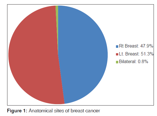
Age distribution of BRCA is shown in Table 1. The patients’ age range was 26-83 years. The peak age incidence for BRCA and their variants were 40-49 years. This accounted for (n = 94/261; 36%). About two-third of all BRCA (n = 159/261; 60.9%) were seen in the 4th and 5th decades of life. Only/261 cases constituting 1.5% were recorded in the 9th decades of life. No case was seen in the in the first and second decades of life. The mean age of was 46 years ± 6.2 S.D. Out of the 261 breast biopsies the left breast accounted for (n = 134/261; 51.3%), the right breast constituted (n = 125/261; 47.9%) while the remaining two cases constituting 0.8% were bilateral breast masses. [Figure 1] Staging at presentation are shown in Table 2. Early presentation (Stages I and II) constituted (n = 62/261; 23.8%) while late presentation (Stages III and IV) accounted for (199/261; 76.2%).
| Age | Frequency (%) |
|---|---|
| 20-29 | 13 (5.0) |
| 30-39 | 65(24.9) |
| 40-49 | 94 (36) |
| 50-59 | 38(14.6) |
| 60-69 | 31(11.9) |
| 70-79 | 16 (6.1) |
| 80-89 | 4(1.5) |
| Total | 261 (100) |
Table 1: Age and frequency distribution of breast cancer
| Stage | Total (%) |
|---|---|
| I | 12 (4.6) |
| II | 50 (19.2) |
| III | 98 (37.5) |
| IV | 101 (38.7) |
| Total | 261 (100) |
Table 2: Stage of breast cancer at presentation
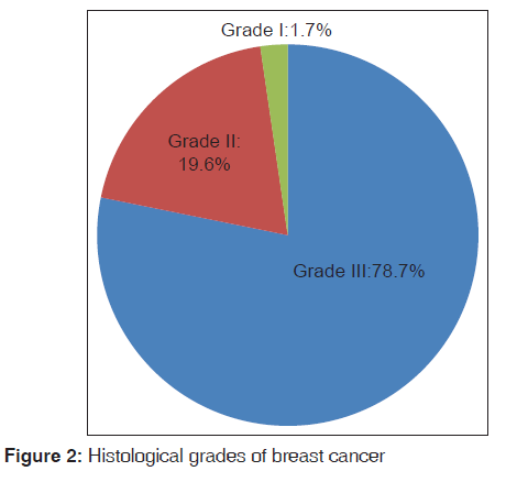
Table 3 shows histological types of BRCA. In this study, invasive carcinoma of no special type (NST) was the most commonly encountered class of carcinoma constituting (n = 203/261, 77.7%) of all BRCA. Of the remaining categories, invasive carcinoma of a special type was (n = 27/261; 10.2%) and carcinoma-in situ constituted (n = 21/261; 8%) while the miscellaneous group of BRCA were relatively rare (n = 11/261; 4.2%). Overall, among all BRCA invasive ductal carcinoma was the most common cancer constituting (n = 183/261; 70%). Invasive lobular carcinoma came a distance second majority accounting for (n = 19/261; 7.2%). The rest of the rare cases are outlined in Table 3. Out of the 261 cases, 240 were graded using the Nottingham grading system. Among these 240 cases, 189 accounting for 78.7% were Grade III while Grade II and I constituted (n = 47/240; 19.6% and n = 4/240; 1.7%) respectively as shown in Figure 2.
| Subtypes | Frequency (%) |
|---|---|
| In situ carcinoma | |
| Lobular | 6(2.3) |
| Ductal | 15 (5.7) |
| Invasive carcinoma (no. special type) | |
| Ductal | 183 (70.0) |
| Lobular | 19 (7.2) |
| Mixed ductal and lobular | 1(0.4) |
| Invasive carcinoma (special types) | |
| Papillary | 7(2.6) |
| Medullary | 4(1.5) |
| Apocrine | 2(0.8) |
| Mucinous | 5(1.9) |
| Clear cell | 3(1.1) |
| Cribriform | 2(0.8) |
| Tubular | 1(0.4) |
| Paget disease | 3(1.1) |
| Miscellaneous (invasive) | |
| Anaplastic | 3(1.1) |
| Metaplastic/carcinosarcoma | 2(0.8) |
| Malignant phylloides tumor | 5(1.9) |
| Pleomorphic sarcoma | 1(0.4) |
| Total | 261 (100) |
Table 3: Histological subtypes of breast cancer
Discussion
Despite the fact that BRCA is the most common cancer in females globally, there are ethnic and geographical variations in its age distribution. In our local environment in Nigeria, patients generally present at a much younger age group when compared with their Caucasian counterparts. In this study, we observed that BRCA occurred most commonly between the age group brackets of 40 and 49 years. This accounted for 36% of all BRCA cases. Again, almost two-third (61%) of all BRCA patients was diagnosed between the 4th to 5th decades. Based on this finding, it could be asserted that the majority of BRCA are seen in pre-menopausal women in our environment. This finding is in tandem with studies by Aftab and Rashid[9] in Pakistan, Asia where BRCA was found the most commonly between 30 and 50 years. Furthermore, similar reports documented by researchers of Sub-Saharan African have also clearly shown that most BRCA cases in Africa are found most commonly in the 4th and 5th decades of life.[10,11] Specifically studies from Cameroun by Kemfang Ngowa et al. reported that 66% of patients with BRCA occurred below 50 years and were all pre-menopausal and perimenopausal women.[12] Yet again, in India subcontinent, the average peak age of occurrence was comparable with what obtains in Sub-Saharan Africa. However, this finding is completely a variance with reports from the United States and European series where most patients with BRCA occurred in post-menopausal women with a peak age incidence in the 7th decades of life.[13] Once more, in the United States whites, the average peak age of BRCA occurrence was 61 years.[13]
The reason for this early age of occurrence of BRCA in this study and other African series is not certain. Nevertheless, it can partly be attributed to low life expectancy in Nigeria and other African countries. It is obvious that Nigerian and indeed African women may not live long enough to present with post-menopausal BRCA. Whether or not, this finding is a true reflection of increase incidence of BRCA in pre-menopausal women in our environment, is a question that needed to be further researched. Once more, high parity among Nigerian and African women have also increase the risk of BRCA before the age of 45 years.[13]
Interestingly, studies have indicated that both African-American and black women in the United Kingdom have similar age of presentation of BRCA to native African women. In spite of the better health-care and higher life expectancy, most cases of BRCA in them present at pre-menopausal and perimenopausal similar to what obtained in African women. This similarity may not be unconnected to genetics involving the BRCA one and two genes in blacks.[14] Furthermore, it has been suggested that blacks women particularly Africans have higher levels of estrogen exposure, which pre-disposes them to higher rates of cell division and deoxyribonucleic acid copying error, resulting eventually to higher risk of early cancer cases.
Our study revealed that late presentation (Stages III and IV) constituted 76.2%. This finding is similar to reports of other researchers. Osime and Dongo[15] and Anyanwu et al.[16] reported 67% and 64% respectively. Adesunkanmi et al.[17] observed that 74% of BRCA present in Stages III and IV. The reason for the late presentation is partly attributable to poverty and ignorance as most cases of late presentations are seen in developing countries including Nigeria.
Histomorphological patterns of BRCA as an important prognostic factor have been well documented. Studies have shown that patients with invasive ductal carcinoma (NST) have a poorer prognosis when compared with other types of BRCA. In this study, invasive ductal carcinoma of NST was the most prevalent histopathological type encountered. This accounted for 70% of all BRCA cases. This finding is similar to other findings in Nigeria. Ekanem and Aligbe in Benin-city reported that invasive ductal carcinoma constituted 75% of all BRCA cases.[18] Similarly, Dauda et al. reported that 78.8% of all BRCA was invasive ductal carcinoma.[19]
Furthermore, similar studies from India reported that invasive ductal carcinoma constituted 88% of all BRCA cases.[3] This is further supported by other reports globally.[20,21] Reports from United States Caucasian series also have it that invasive ductal carcinoma was also the most common BRCA.[20] In our study, invasive lobular carcinoma occurred a distance second. This constituted 7.2% of all cases. This again, indeed is comparable with a similar reports by Dauda et al. and Nggada et al. where it constituted 6.7% and 6.6% respectively.[19,21] This yet again is at variance with studies done in the United States invasive lobular carcinoma accounted for 15%.[20,21] The reason for this variation is partly due to the fact that in the United States all H and E slides are further re-confirmed with immunohistochemistry. In this study, intraductal carcinoma accounted for 5.7% of all BRCA cases. Similar report by Nggada et al. in Maiduguri, North-Eastern Nigeria and Kene et al. in Zaria, North-Western Nigeria revealed that intra-ductal carcinoma constituted 6% and 3% respectively.[21,22] Nevertheless a very high incidence of 20% of intra-ductal carcinoma was documented in the United State.[23] The reason for this marked variation is attributed to very efficient screening programs and high level of awareness of BRCA with subsequent early presentation in the United States.
In this study, the left breast with the upper outer quadrant as the most commonly affected anatomical sites accounting for 51.3% and closely followed by the right breast accounting for 47.9% and the least common was bilateral breast tumor constituting 0.8%. From this observation, unilateral breast lesions therefore constituted 99.2%. This is similar to other studies done by Saxena et al.[3] in India and Ekanem and Aligbe[18] in Benin, where unilateral breast lumps accounted for 99.0%, 99.2% respectively. This again is similar to work carried out by other researchers locally and globally.
In general, studies have it that, histological Grade III breast carcinoma characterized by high grade nuclear atypia, extensive necrosis and increase number of positive nodes is more common in African women than Caucasians. More specifically in Nigeria and Tanzania, majority of BRCA constituting 56% and 45% have Grade III breast carcinoma.[11,24] This is completely at variance with reports from Finland where histological Grade III BRCA constituted only 16%.[21] Once more, studies confirm the similarities of histological grade of BRCA in black British and African-American women to their African counterparts.[24] This is partly due to the reason why the BRCA progresses more aggressively in black women as compared with their caucasian counterparts.
One major challenge encountered in this study is the follow-up to management of patients. Most of the patients may not have come back for follow-up biopsies for histology after initial treatment with surgery, chemotherapy, radiotherapy and hormone therapy. This is similar to observations of other researchers.[15-17] The reasons partly attributed to this observation is based on the fact that some patients may have being referred to other centers with immunohistochemistry facilities and hormone therapy for further management. Again some of the patients with advanced stages of the disease may have died in the process of looking for a cure. Still financial constraint, poverty and search for alternative medication may also influence the follow-up of these patients.
Limitations
The cases in this study did not have immunohistochemistry done to determine the hormone receptor status. The reason for this is due to the fact that this center has various challenges in carrying out immunohistochemistry. Hormone receptor analysis is therefore not part of the result analyzed.
Conclusion
Nearly two-third (61%) of all BRCA patients was diagnosed in the 4th and 5th decades of life. Again, at least nine out of every 10 cases of BRCA in our environment are invasive with majority as invasive ductal carcinoma of NST and majority presenting as Grade III tumors. In our environment most cases present late due to poverty, lack of awareness, alternative medication and psychological fear that mastectomy may interfere with their womanhood.
Source of Support
Nil.
Conflict of Interest
None declared.
References
- Arab SM. Medicine in Ancient Egypt, part 2 of 3. Available from: http://www.arabworldbooks.com/articles 8b.htm. [Accessed on 2007 May 3].
- Ferlay J, Shin H, Bray F, Mathers C, Forman D, Parkin DM. Globocan 2008 v1.2, Cancer Incidence and Mortality Worldwide. IARC Cancerbase No 10. Lyon, France: International Agency for Research on Cancer; 2010. Available from: http://www.globocan.iarc.fr/. [Accessed on 2012 August].
- Saxena S, Rekhi B, Bansal A, Bagga A, Chintamani, Murthy NS. Clinico-morphological patterns of breast cancer including family history in a New Delhi hospital, India – A cross-sectional study. World J Surg Oncol 2005;3:67.
- Anderson WF, Devesa SS. In situ male breast carcinoma in the surveillance, epidemiology, and end results database of the National Cancer Institute. Cancer 2005;104:1733-41.
- Althuis MD, Dozier JM, Anderson WF, Devesa SS, Brinton LA. Global trends in breast cancer incidence and mortality 1973-1997. Int J Epidemiol 2005;34:405-12.
- American Cancer Society. Cancer Facts and Figures 2010. Atlanta: American Cancer Society; 2010.
- Bhurgri Y, Kayani N, Faridi N, Pervez S, Usman A, Bhurgri H, et al. Patho-epidemiology of breast cancer in Karachi ‘1995-1997’. Asian Pac J Cancer Prev 2007;8:215-20.
- Travassoli FA, Deville P, editors. World Health Organization Classification of Tumours: Pathology and Genetics of Tumours of the Breast and Female Genital Organs. Lyon: IARC Press; 2003.
- Aftab ML, Rashid A. A clinicopathological study of carcinoma breast. Pak J Health 1998;35:96-8.
- Gukas ID, Jennings BA, Mandong BM, Igun GO, Girling AC,Manasseh AN, et al. Clinicopathological features and molecular markers of breast cancer in Jos, Nigeria. West Afr J Med 2005;24:209-13.
- Gakwaya A, Kigula-Mugambe JB, Kavuma A, Luwaga A, Fualal J, Jombwe J, et al. Cancer of the breast: 5-year survival in a tertiary hospital in Uganda. Br J Cancer 2008;99:63-7.
- Kemfang Ngowa JD, Yomi J, Kasia JM, Mawamba Y, Ekortarh AC, Vlastos G. Breast cancer profile in a group of patients followed up at the radiation therapy unit of the Yaounde General Hospital, Cameroon. Obstet Gynecol Int 2011;2011:143506.
- Bowen RL, Duffy SW, Ryan DA, Hart IR, Jones JL. Early onset of breast cancer in a group of British black women. Br J Cancer 2008;98:277-81.
- Gao Q, Neuhausen S, Cummings S, Luce M, Olopade OI. Recurrent germ-line BRCA1 mutations in extended African American families with early-onset breast cancer. Am J Hum Genet 1997;60:1233-6.
- Osime CO, Dongo AE. Presentation, pattern and outcome of breast cancer in a poor economy: A definition of the tripod of ignorance, disease and poverty. Emerg Med J 2012;11:20-8.
- Anyanwu SN, Egwuonwu OA, Ihekwoaba EC. Acceptance and adherence to treatment among breast cancer patients in Eastern Nigeria. Breast 2011; 20 Suppl 2:S51-3.
- Adesunkanmi AR, Lawal OO, Adelusola KA, Durosimi MA. The severity, outcome and challenges of breast cancer in Nigeria. Breast 2006;15:399-409.
- Ekanem VJ, Aligbe JU. Histopathological types of breast cancer in Nigerian women: A 12-year review (1993-2004). Afr J Reprod Health 2006;10:71-5.
- Dauda AM, Misauno MA, Ojo EO. Histopathological types of breast cancer in Gombe, North Eastern Nigeria: A seven-year review. Afr J Reprod Health 2011;15:109-11.
- Li CI, Anderson BO, Daling JR, Moe RE. Trends in incidence rates of invasive lobular and ductal breast carcinoma. JAMA 2003;289:1421-4.
- Nggada HA, Yawe KD, Abdulazeez J, Khalil MA. Breast cancer burden in Maiduguri, North Eastern Nigeria. Breast J 2008;14:284-6.
- Kene TS, Odigie VI, Yusufu LM, Yusuf BO, Shehu SM, Kase JT. Pattern of presentation and survival of breast cancer in a teaching hospital in North Western Nigeria. Oman Med J 2010;25:104-7.
- Burstein HJ, Polyak K, Wong JS, Lester SC, Kaelin CM. Ductal carcinoma in situ of the breast. N Engl J Med 2004; 350:1430-41.
- Ikpat OF, Kronqvist P, Kuopio T, Ndoma-Egba R, Collan Y. Histopathology of breast cancer in different population. Comparative analysis for Finland and Africa. Electron J Pathol Histol 2002;8:24-31.


 The Annals of Medical and Health Sciences Research is a monthly multidisciplinary medical journal.