Inter-Canthal and Inter Alar Distance as a Predictor of Width of Maxillary Central and Lateral Incisor – An In Vivo Study
2 Department of Maxillofacial Prosthodontics and Implantology, Peoples Dental Academy, Bhopal, Madhya Pradesh, India, Email: naveenyadav_s@gmail.com
3 Department of Maxillofacial Prosthodontics and Implantology, Peoples College of Dental Sciences & Research Centre, Bhopal, Madhya Pradesh, India, Email: sunilmsr200@yahoo.co.in
Citation: Dwivedi A, Yadav NS, Mishra SK. Inter-Canthal and Inter Alar Distance as a Predictor of Width of Maxillary Central and Lateral Incisor –An In Vivo Study. Ann Med Health Sci Res. 2017; 7:276-279
This open-access article is distributed under the terms of the Creative Commons Attribution Non-Commercial License (CC BY-NC) (http://creativecommons.org/licenses/by-nc/4.0/), which permits reuse, distribution and reproduction of the article, provided that the original work is properly cited and the reuse is restricted to noncommercial purposes. For commercial reuse, contact reprints@pulsus.com
Abstract
Background: The solution and arrangement of maxillary anterior teeth for edentulous patients in a natural and aesthetically pleasing form has remained an elusive and challenging endeavour. Aim: The aim of the study was to evaluate co-relation between maxillary right & left central and lateral incisors with inter canthal distance and between maxillary right and left central and lateral incisors with inter alar width. This study was done to determine whether inter-canthal distance and inter-alar width can be used as a predictor of width of maxillary central and lateral incisors. Subjects and Methods: To evaluate the relationship of inter canthal distance and inter alar width with the maxillary central incisor and maxillary lateral incisor, measurements from 312 subjects were obtained. Their age ranged from 18-24 years. This study measured and calculated the inter canthal distance from the medial angle to the medial angle of the palpebral fissure, width of the nose also measured and compared with the width of maxillary right and left central and lateral incisors. The mean, standard deviation, maximum and minimum values were calculated. Karl Pearson’s correlation coefficient and p-value were calculated for significance. Results: There was no significant correlation between Inter canthal distance with Right central incisor width, Right lateral incisor width, Left central incisor width & Left lateral incisor width but there was significant correlation between Inter-alar width with Right central incisor width, Right lateral incisor width & Left central incisor width. Conclusion: Inter-alar width can be used as a preliminary method for determining the width of the maxillary right and left central incisors and maxillary right lateral incisor for edentulous patients.
Keywords
Central incisor width; Inter-alar width; Inter Canthal distance; Lateral incisor
Introduction
Everyone is concerned about their facial appearance as it is a very significant part of self-image [1]. The artisans of the dental profession since many years had suggested different norms, guidelines and criteria for aesthetic selection of teeth and their arrangement. The appearance of the teeth within the framework of the face decides the success of an aesthetically sound denture [2]. However for the edentulous patients, it remained an elusive and challenging endeavour to select and arrange maxillary anterior teeth in a natural and aesthetically pleasing form [3].
Various efforts had been made since long but no universally acceptable method has been established to meet this end. To attain acceptable results, using their clinical expertise and aesthetic sense dentist seeks guidance from various techniques. It remains a prime concern for the Prosthodontist to follow the rationale or criteria that should be used for selection and arrangement of artificial teeth [4].
Different guidelines have been given for determining the width of the maxillary anterior teeth when pre-extraction records are not available. These guides include bizygomatic width [5]. canthus distance [6]. inter pupillary width [7]. interalar width [8]. Different views and conflict had been reported on the significance of the interalar width in the selection of anterior teeth [9,10].
When anterior teeth are selected for edentulous patients, the mesiodistal width of maxillary central incisors and maxillary lateral incisors are important because they are the most prominent teeth in the arch [1].
The maxillary anterior teeth are the key elements contributing to the aesthetic importance of what we call dentofacial beauty. It’s our duty as dentist to preserve the natural dignity of advancing age while fabricating prosthesis, with appropriate and careful selection and arrangement of teeth. Hence this study was conducted to determine the inter-canthal distance and inter- alar width for selection of maxillary central and lateral incisor.
Subjects and Methods
The present study was conducted in the Department of Prosthodontics, Crown and Bridge, People’s Dental Academy, Bhopal, Madhya Pradesh, India from March 2013 to August 2013. The sample sizes were randomly selected from the outpatient department keeping power of the study at 80%. Three hundred and twelve subjects were selected for the study; all subjects were between 18-24 years. Subjects presented with full complement of natural teeth and having intact contact, without having any Orthodontic or Prosthodontic treatment and no abnormal or altered nose. The purpose of this study was to evaluate the usefulness of inter-canthal distance and interalar width in selection of maxillary central and lateral incisor width. The subjects were selected following the inclusion and exclusion criteria. An ethical clearance was obtained from the ethics committee of the university and an informed consent was obtained from the study subjects. Two investigators measured three parameters independently for each subject three times to get as much as accurate mean value.
Inter Canthal Distance (ICD) Measurement
Subjects were seated with their heads supported in an upright position and they looked straight. The sterilized caliper was placed against the forehead and lowered toward the eyes. The arms of the calipers were adjusted so that they were in gentle contact with the medial angle of the palpebral fissures of the eyes. Utmost care was taken not to compress the soft tissue. The distance between these two anatomical landmarks was recorded as ICD to an accuracy of 0.01 mm. Inter-canthal distance was measured three times for each subject by the same operator. Average value was taken to avoid intra-operator observational errors.
Determination of Inter-Alar Width (IAW)
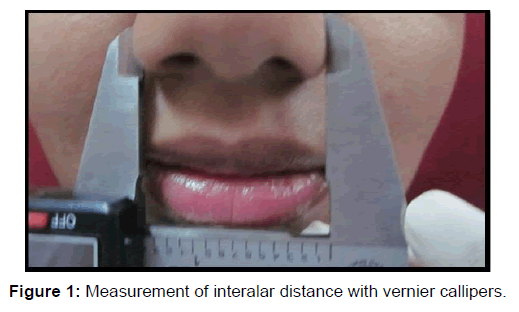
The inter-alar width was determined by using the external width of the nose at the widest point. The distance between these two points was measured using a digital vernier caliper without the application of pressure by bringing the recording parts of the caliper just in contact with the outer surface of the nose [Figure 1]. While measuring, the patient was asked to stop breathing momentarily to avoid any change in shape of the nose. Each measurement was mean of three readings.
Central Incisor Width (CIW) and Lateral Incisor Width (LIW)
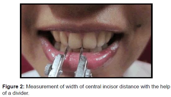
Maxillary central incisors and lateral incisors were measured mesiodistally at their contact point with the help of a divider. This divider could be fixed in position with a screw thread and it had finely pointed ends that fitted interdentally accurately [Figure 2]. Each tooth was measured three times by the same operator and the average was taken. This was done to ensure observational reliability of the procedure.
For all the subjects in the study mean, standard deviation, maximum and minimum values were calculated. Karl Pearson’s correlation coefficient and p-value were calculated for significance.
Karl Pearson’s Correlation Coefficient shows the relationship between two values. It is expressed as a single figure. It is calculated so that its numerical value is between +1 and -1. A positive value means that as one measurement increases in value, the other one also increases in value. A minus value means that as one measurement increases in value, the other one decrease.
All statistical analyses were done with the help of the statistical package for social scientists (SPSS Version 16; Chicago Inc., USA) computer software for Windows version 7.
Results
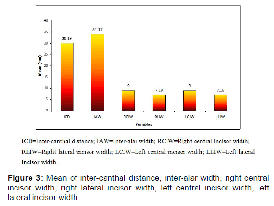
The mean, standard deviation and p value for Inter canthal distance, Inter alar width, Right central incisor width (RCIW), Left central incisor width (LCIW), Right lateral incisor width (RLIW), Left lateral incisor width (LLIW) were given in Tables 1 and 2 and Figure 3. There was no significant correlation between Inter canthal distance with Right central incisor width, Right lateral incisor width, Left central incisor width & Left lateral incisor width. There was significant correlation between Inter-alar width with Right central incisor width, Right lateral incisor width & Left central incisor width.
| Main | Group | Mean | SD | N | r | p value |
|---|---|---|---|---|---|---|
| Inner Canthal distance | ICD | 30.2 | 2.6 | 312 | 0.09 | 0.12 |
| RCIW | 9.00 | 0.5 | 312 | |||
| ICD | 30.2 | 2.6 | 312 | 0.05 | 0.44 | |
| RLIW | 7.2 | 0.6 | 312 | |||
| ICD | 30.2 | 2.6 | 312 | 0.04 | 0.47 | |
| LCIW | 9.00 | 0.5 | 312 | |||
| ICD | 30.2 | 2.6 | 312 | 0.09 | 0.11 | |
| LLIW | 7.2 | 0.5 | 312 |
P<0.05 was significant
ICD=Inter-canthal distance; RCIW= Right central incisor width; LCIW=Left central; incisor width; RLIW= Right lateral incisor width; LLIW= Left lateral incisor width; N=Number of subjects; r= Correlation coefficient.
Table 1: Relationship between inter-canthal distances with other variables.
| Main | Group | Mean | SD | N | r | p value |
|---|---|---|---|---|---|---|
| Inter alar width | IAW | 34.17 | 2.76 | 312 | 0.176 | P<0.01 |
| RCIW | 9.00 | 0.54 | 312 | |||
| IAW | 34.17 | 2.76 | 312 | 0.128 | P<0.01 | |
| RLIW | 7.19 | 0.58 | 312 | |||
| IAW | 34.17 | 2.76 | 312 | 0.166 | P<0.01 | |
| LCIW | 9.00 | 0.53 | 312 | |||
| IAW | 34.17 | 2.76 | 312 | 0.074 | 0.19 | |
| LLIW | 7.18 | 0.53 | 312 |
P<0.05 was significant
IAW=Inter-alar width; RCIW= Right central incisor width; LCIW=Left central; incisor width; RLIW= Right lateral incisor width; LLIW= Left lateral incisor width. N=Number of subjects; r= Correlation coefficient
Table 2: Relationship between inter-alar width with other variables.
Discussion
While numerous methods have been suggested for estimating the combined width of the maxillary anterior teeth and the central incisor, there seem to be few reliable guidelines and many conflicting views.[3-10] In the construction of complete dentures, the estimation of the combined width of maxillary six anterior teeth is an important clinical procedure when preextraction records are not available.
When anterior teeth are selected for edentulous subjects, the mesiodistal width of the maxillary central incisors is important because they are the most prominent teeth in the arch when viewed from the frontal aspect. Shillingburg et al. reported that the combined width of the maxillary central incisors occupied 37% of the circumferential arch distance between the distal surface of the maxillary canines. The combined width of the lateral incisors and canines accounted for 31% and 32% of the distance, respectively [11].
Authors have suggested that the width of the nose serves as a guide for the selection of width of maxillary anterior teeth. They stated that “parallel” lines extended from the lateral surface of the alae of the nose onto the labial surface of occlusal rim could be used to estimate the position of maxillary canine [12]. According to the embryogenic philosophy, the nose has been considered as an essential guide for the selection of the size of the upper incisors. The nose and the four upper maxillary incisors develop from the same embryonic origin called the fronto nasal process. Furthermore the interalar width is a facial landmark that is at the closest distance from the teeth [13].
It is a moral responsibility of Prosthodontist to preserve the natural dignity of advancing age while fabricating prosthesis, with appropriate and careful selection and arrangement of teeth. Further demographic variation may exist with repeat to anthropometric measurements [4]. This study was conducted to determine the relationship of the inter canthal distance (ICD) and Inter-Alar width (IAW) with the width of Maxillary right and left Central Incisors and Lateral Incisors.
The mean value of Inter-Canthal distance in present study was 30.19 mm. Deogade et al. [14]. in their study on Indian population found the mean value of inter canthal distance as 28.04 mm in males and 24.4 mm in females. The mean inter canthal distance in this study was lesser than the value reported by Esan et al. as 36.1 mm in the Nigerian population [15]. The measurements being recorded in different populations and of different countries might be the reason for variation in the value. The mean Inter- Alar width was 34.17 mm. This distance was lesser than the value reported by Miranda et al. as 38.52 mm [8].
However, this study has shown that this approach cannot be applied as a gold standard in all cases. From the current study it may be stated that a proportional relationship exists between the widest part of the nose and the front of the dental arch as was presented in past studies. This study brings to light that the Inter- Alar width and the width of maxillary right and left central and lateral incisors are not always equivalent. However there is sufficient correlation to use the Inter-Alar width to obtain the approximate width of maxillary right and left central and lateral incisors while selecting the maxillary anterior teeth for complete dentures.
The limitation of this study was resiliency of the soft tissue. Further this study was done within the institutional set up and subjects of the age group between 18 to 24 years were only evaluated. Hence the results may be applicable to just a small population in the said age range. The study can be more complete if variety of age and variety of Indian sub population groups are considered. The result of the study should be validated including a large population size spread over the entire Indian subcontinent. This would help to generate multiplication factor for various anthropological measurements for use limited to the Indian population.
Conclusion
There was significant correlation found between inter-alar width with maxillary right and left central incisors and maxillary right lateral incisor. Hence it can be concluded that inter alar width is a good predictor of width of maxillary right and left central incisor and maxillary right lateral incisor whereas inter canthal distance is not at all a good predictor for width of maxillary central and lateral incisors.
Conflict of Interest
To the best of our knowledge no conflict of interest, financial or other, exists.
REFERENCES
- George S, Bhat V. Inner canthal distance and golden proportion as predictor of maxillary central incisor width in South Indian population. Indian J Res 2010;21:491-495.
- Bolla SC, Gantha NS, Sheik RB. Review of history in the development of esthetics in dentistry. J Dent Med Sci 2014;13:31-35.
- Debnath N, Gupta R, Meenakshi A, Kumar S, Hota S, Rawat P. Relationship of inter-condylar distance with inter-dental distance of maxillary arch and occlusal vertical dimension: A clinical anthropometric study. J Clin Diagn Res 2014;8:ZC39-ZC43.
- Patel JR, Sethuraman R, Naveen YG, Shah MH. Comparative evaluation of the relationship of inner-canthal distance and inter-alar width to the inter-canine width amongst the Gujarati population. J Adv Oral Res 2011;2:31-38.
- Baleegh S Choudhry Z, Malik S, Baleegh H. The relationship between widths of upper anterior teeth and facial widths. Pak Oral Dent J 2015;35:742-745.
- Ellakwa A, McNamara K, Sandhu J, James K, Arora A, Klineberg I, El-Sheikh A, Martin FE. Quantifying the selection of maxillary anterior teeth using intraoral and extraoral anatomical landmarks. J Contemp Dent Pract 2011;12:414-421.
- Cesario VA, Latta GH. Relationship between the mesiodistal width of the maxillary central incisor and interpupillary distance. J Prosthet Dent 1984;52:641-643
- Miranda GA, D'Souza M. Evaluating the reliability of the interalar width and intercommissural width as guides in selection of artificial maxillary anterior teeth: A clinical study. J Interdiscip Dentistry 2016;6:64-70.
- Gomes VL, Goncalves LC, Costa MM, Lucas BL. Interalar distance to estimate the combined width of the six maxillary anterior teeth in oral rehabilitation treatment. J Esthet Restor Dent 2009;21:26‑35.
- Sinavarat P, Anunmana C, Hossain S. The relationship of maxillary canines to the facial anatomical landmarks in a group of Thai people. J Adv Prosthodont 2013;5:369‑373.
- Shillinberg HT, Kaplan MJ, Grace CS. Tooth dimensions - A comparative study. J South Calif Dent Assoc 1972;40:830-839.
- Rai R. Correlation of nasal width to inter-canine distance in various arch forms. J Indian Prosthodont Soc. 2010;10:123-127.
- Sülün T, Ergin U, Tuncer N. The nose shape as a predictor of maxillary central and lateral incisor width. Quintessence Int 2005;36: 603-607.
- SC, Mantri SS, Sumathi K, Rajoriya S. The relationship between innercanthal dimension and interalar width to the intercanine width of maxillary anterior teeth in central Indian population. J Indian Prosthodont Soc. 2015;15:91-97.
- Esan TA, Oziegbe OE, Onapokya HO. Facial approximation: Evaluation of dental and facial proportions with height. Afr Health Sci 2012;12:63–68.


 The Annals of Medical and Health Sciences Research is a monthly multidisciplinary medical journal.