K.S. Micro‑Implant Placement Guide
- *Corresponding Author:
- Archana Damle
Dawn W Foster Psychiatry Department at Yale Schol of Medicine Yale University, New Haven, CT 06519, USA Tel: 203 974 7892
E-mail: drasangwan@gmail.com
Abstract
A one of the greatest concerns with orthodontic mini-implants is risk of injury to dental roots during placement is, especially when they are inserted between teeth. Many techniques have been used to facilitate safe placement of interradicular miniscrews. Brass Wires or metallic markers are easy to place in the interproximal spaces, but because their relative positions may be inconsistent in different radio -graphic views, they are not always accurate. K.S. micro implant placement guide suggested in this article is simple design and easy in fabrication, required minimal equipment for fabrication and does not disturb the existing appliance system, clearly located in the radiograph and the mini-screw can be easily inserted through the guide reducing the chance of implant misplacement.
https://betkolikgirisi.com https://betlikeguncel.com https://betparkagiris.com https://bettickett.com https://betturkeyegiris.com https://extrabetgirisi.com https://holiganbeti.com https://ilbete.com https://ikimisligirisi.com https://imajbetegir.com https://jojobeti.com https://kralbetting.com https://mariogiris.com https://marsbahise.com https://meritegiris.com https://milanobeti.com https://piabetegir.com https://redwinegiris.com https://supertotobete.com https://tempobetegir.com
Keywords
Orthodontic, Mini-implants, microimplant jig, microimplant guide, microimplant positioning, miniscrew
Introduction
Mini-screws offer skeletal anchorage and having many clinical uses and advantages, available in varied sizes and shapes, there are various sites for placement according to uses. Micro-implants are ease of insertion and removal, the ability to load forces immediately and rapid healing. A one of the greatest concerns with orthodontic mini-implants is a risk of injury to dental roots during placement is, especially when they are inserted between teeth.[1] Placement of a mini-screw too close to the root can also result in insufficient bone remodeling around the screw and transmission of occlusal forces through the teeth to the screw, which can lead to implant failure.[2] Even though, periodontal structures can heal after being injured by temporary orthodontic anchorage devices,[3] it is important to carefully select insertion sites using the clinical and radiographic evaluation of their anatomical details.
Many techniques have been used to facilitate safe placement of interradicular mini-screws. Brass wires[4] or metallic markers[5] are easy to place in the interproximal spaces, but because their relative positions may be inconsistent in different radiographic views, they are not always accurate. K.S. micro-implant placement guide suggested in this article is a simple design and easy in fabrication, required minimal equipment for fabrication and does not disturb the existing appliance system, clearly located in the radiograph and the mini-screw can be easily inserted through the guide reducing the chance of implant misplacement.[6]
K.S. Micro-Implant Placement Guide Fabrication
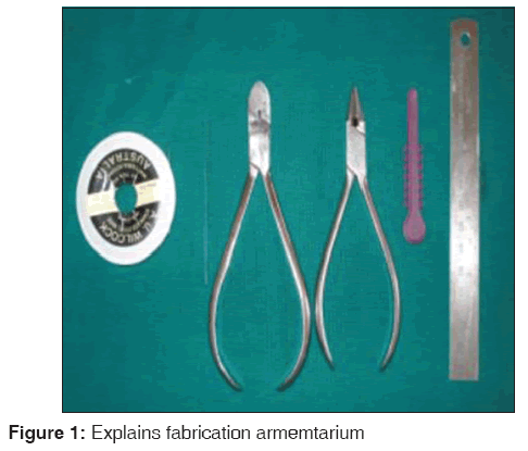
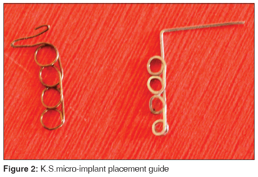
The wire guide is fabricated using round 0.018 or 0.020 (A.J. Wilcock) or 17 × 25 or 19 × 25 stainless steel wire [Figure 1]. A helix of 2–3 mm diameter is made at the center of the wire [Figure 2]. The appropriate length is determined by the desired mini-screw insertion point (generally 5–6 mm apical to the alveolar crest). After vertical height is determined, continues vertical loop made until measured length and one or two horizontal bends are the place at the level of the adjacent brackets.
Here, we are using multiple loops helix as they are helpful in area determination as compared to single helix and also if we use thickened wire than stability will be more, as the gap between the slots will be filled.
Clinical Procedure
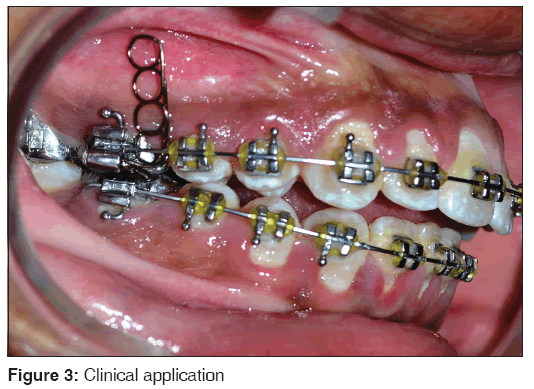
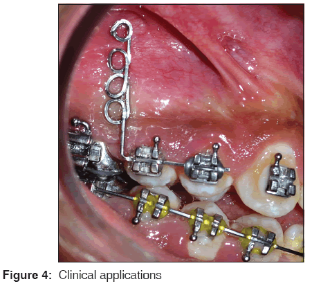
The wire guide is secured with a bracket or tube by ligature or an “O” ring. The mini-screw is inserted through the helix of the guide in the desired direction. The wire guide is disengaged after three-fourth of the mini-screw is driven in and then the mini-screw is completely inserted. Placement accuracy is reconfirmed clinically [Figures 3 and 4]. A periapical radiograph is taken to confirm the correct position of the helixes for the mini-screw insertion the intraoral periapical radiograph here shown was taken by cone-beam paralleling technique.
Discussion
The use of mini-implants in orthodontics has phenomenally increased in the last few years due to the clear advantage of their clinical efficiency in anchorage control.
The critical issue about mini-implants is the location of the implant. It is very important to place the implant in a location that does not damage adjacent roots or other vital tissues. There are various methods mentioned in the literature; e.g. using a mesh placed in an interradicular area and radiographed to use as a guide to implant placement, or custom-made soldered wire guides placed both buccally and lingually and then radiographed to use as a guide.
The technique mentioned in the present article is simple and accurate because the implant is inserted through the helix of the guide, hence less chance for it to be misplaced. The existing orthodontic appliance is not disturbed and the implant guide can be disengaged without any difficulty.
We can also use rectangular wire and we can place one loop over another to increase the thickness so that angulation does not get changed. Here reason for using wire. 018 is because if we use. 014 the stability will get compromised.
Conclusion
K.S. micro-implant placement guide is simple in design, easy to fabricate, inexpensive, supportive and can be used with a variety of mini-screws. Here, we used 0.018 wire as it engages the bracket slots properly hence giving better stability. Further we are researching on it for betterment like changing the gauge and increasing the loop thickness. However, still lots of modifications are yet to be searched.
References
- Choi HJ, Kim TW, Kim HW. A precise wire guide for positioning interradicular miniscrews. J Clin Orthod 2007;41:258-61.
- Kuroda S, Yamada K, Deguchi T, Hashimoto T, Kyung HM, Takano-Yamamoto T. Root proximity is a major factor for screw failure in orthodontic anchorage. Am J Orthod Dentofacial Orthop 2007;131:S68-73.
- Asscherickx K, Vannet BV, Wehrbein H, Sabzevar MM. Root repair after injury from mini-screw. Clin Oral Implants Res 2005;16:575-8.
- Kyung HM, Park HS, Bae SM, Sung JH, Kim IB. Development of orthodontic micro-implants for intraoral anchorage, J. Clin. Orthod. 37:321-328, 2003.
- Carano A, Velo S, Leone P, Siciliani G. Clinical applications of the Miniscrew Anchorage System, J. Clin. Orthod. 39:9-24, 2005.
- Ludwig B, Glasl B, Bowman SJ, Wilmes B, Kinzinger GS, Lisson JA. Anatomical guidelines for miniscrew insertion: Palatal sites. J Clin Orthod 2011;45:433-41.


 The Annals of Medical and Health Sciences Research is a monthly multidisciplinary medical journal.