Male Urethral Diverticulum Having Multiple Stones
- *Corresponding Author:
- Dr. Pankaj Kumar Garg,
Department of Surgery, University College of Medical Sciences and Guru Teg Bahadur Hospital, Dilshad Garden, New Delhi - 110 095, India.
E-mail: dr.pankajgarg@gmail.com
Citation: Mohanty D, Garg PK, Jain BK, Bhatt S. Male urethral diverticulum having multiple stones. Ann Med Health Sci Res 2014;4:53-5.
Abstract
Congenital diverticulum of male urethra is an uncommon entity. Neglected management complicates the process in the form of calculi formation and recurrent urinary infection. A 10âГғвҖҡГӮвӮ¬ГғвҖҡГӮвҖҳyearâГғвҖҡГӮвӮ¬ГғвҖҡГӮвҖҳold boy presented with urinary voiding disturbances and development of a painless hard lump at the penoscrotal junction. Imaging demonstrated presence of anterior urethral diverticulum with contained calculi in it. Open urethral diverticulectomy, extraction of multiple calculi, and primary urethral reconstruction over a Foley catheter was carried out. Early diagnosis and individualized surgical management of congenital male urethral diverticulum is the key to a successful outcome.
Keywords
Diverticulum, Stones, Urethra
Introduction
Urolithiasis is a common problem affecting the global population. Lower urinary tract particularly the urethra is an unusual site for urolithiasis accounting for only 0.3% patients of urinary calculi.[1] Forceful jet of voided urine with complete post‑micturition collapse of urethral lumen accounts for this low incidence. The urethral calculi are mostly migratory in nature originating in the urinary bladder and upper urinary tract. Calculi may also develop de novo in association with urethral foreign body and diverticulum. Development of urethral calculi in a pre‑existing diverticulum involving the male urethra is seldom encountered in clinical practice. We herein report a young boy with congenital urethral diverticulum (UD) containing numerous calculi to highlight this unusual occurrence.
Case Report
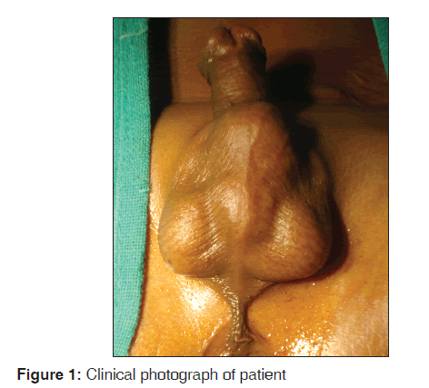
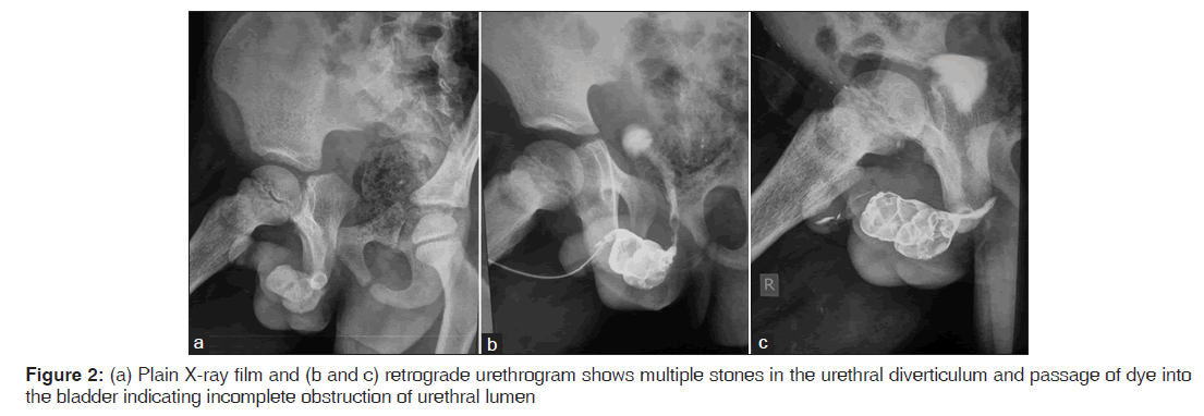
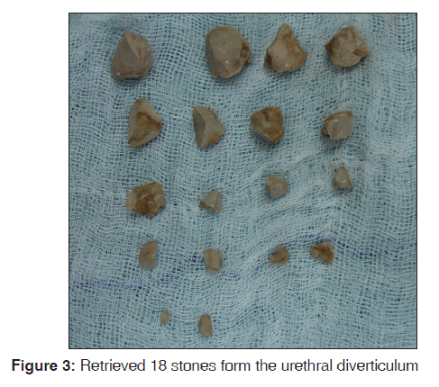
A 10‑year‑old boy presented to us complaining of multiple scrotal swellings and burning during micturation of 3 months duration. He also reported to have occasional painful and difficult urination. He did not complain of hematuria, fever or any bowel complaint. Physical examination revealed multiple non tender and hard scrotal swellings in relation to bulbar urethra [Figure 1]. Ultrasound of the scrotum suggested possibility of large UD with multiple stones. Retrograde urethrogram showed multiple stones in the UD and passage of dye into the bladder indicating incomplete obstruction of urethral lumen [Figure 2]. He underwent urethral diverticulectomy with end‑to‑end urethral anastomosis in view of large size of UD and multiple stones [18 in number, Figure 3]. Post‑operative period was uneventful. He is asymptomatic after 6 months of follow‑up.
Discussion
Urethral diverticulum (UD) is an out‑pouching of the urethral wall and having a free communication with the urethral lumen. The male UD is classified into congenital and acquired types. The congenital diverticulum is the result of segmental defect of the urethral wall due to imperfect closure of urethral folds on the ventral aspect.[2] Anterior urethra, particularly the penoscrotal junction, is the preferred site for development of congenital diverticulum. Congenital diverticulum is characterized by having a true epithelial lining, wall being formed by full thickness urethral musculature, and manifesting early in life. Acquired diverticula are secondary to distal obstruction to urine flow such as hypospadias and urethral stricture, suppuration and necrosis of urethral wall following trauma, instrumentation, and drainage of a prostatic or periurethral abscess. They are lined by granulation tissue, lack smooth muscle fibers in their wall, commonly encountered in adults, and involve the posterior urethra. Differential diagnosis for UD includes syringoceles (cystic dilatation of the Cowper’s gland), sequestration cysts, epidermoid and epithelial inclusion cysts.
Most patients with uncomplicated UD remain asymptomatic for prolonged duration of time. Complications stem from inadequate drainage and commonly manifest as urinary voiding symptoms. Common presentations are thin stream, post void dribbling, recurrent urinary tract infection, painless penoscrotal lump, and rarely extravasations of urine secondary to urethral rupture. The stasis of urine predisposes to infection, crystallization and formation of calculi. Development of calculi is observed in 4‑10% of patients with UD.[3] Since the actual urethral lumen is not completely occluded by the calculi inside the diverticulum, presentation with acute urinary retention (AUR) is rare. In contrast, migratory calculi form upper urinary tract often gets impacted in urethra leading to development of AUR. Presence of pinhole meatus, palpable urethral calculi, defect in continuity of corpus spongiosum and spongiofibrosis on focused local examination of the external genitalia aid in clinical diagnosis of this entity.
Radiological delineation of UD is essential for outlining the treatment modality. Plain roentgenogram of KUB (Kidney, Ureter and Bladder) area is useful to document the presence of calculi in urethra. Demonstration of the UD is facilitated by ascending urethrogram, micturating cystourethrogram and urethrosonography. Both fluoroscopic and ultrasonographic evaluation are complimentary to each other and accurately demonstrate the location and size of the diverticulum, caliber of the neck, and the diameter of the proximal and distal urethra in relation to the diverticulum. Additionally, urethrosonography can provide useful information about the thickness of the wall of the diverticulum and the adjacent urethra that have a significant effect on the choice of surgical procedure. The value of magnetic resonance imaging is proven in female UD and its use is increasing for diagnosis of male UD.[4] A gentle endoscopic evaluation helps in visualization of associated anterior urethral valves, size of the neck of the diverticulum, appearance of the adjacent urethral mucosa and evaluation for feasibility of endoscopic definitive management.
Management of UD is guided by the presenting symptoms and the associated anatomical abnormalities. Small incidentally detected uncomplicated diverticulum can be managed non‑operatively, provided the patient commits to regular follow‑up. The patients are counseled regarding decompression of the UD by external manual urethral pressure following the act of micturition and precaution to prevent urinary tract infection. Complicated diverticula invariably require surgical intervention that can be open or endoscopic. Endoscopic deroofing of the diverticular sac, fulguration of the associated anterior urethral valve, and stone fragmentation by pneumatic or laser lithotripter is an attractive option; however, only a limited number of patients qualify for this treatment modality. Patients with large diverticulum, large stone burden, thick and fibrotic diverticulum wall, deficient corpus spongiosum, and unhealthy adjacent supportive tissue are candidates for open reconstructive surgery. Complete diverticulectomy followed by primary urethral reconstruction allows satisfactory recovery in most of the patients. Primary reconstruction is considered unsafe in the presence of large defect following diverticulectomy, compromised blood supply to the cut ends, tension on suture line, unhealthy urethral mucosa, and heavy bacterial contamination of urine.[5] Reinforcement of repair with genital (penile skin, dartos, tunica vaginalis) or extra genital (buccal mucosa) graft reduces the incidence of post‑operative urethra‑cutaneous fistula in these patients.
In conclusion, young male individuals presenting with voiding disturbances should be thoroughly investigated for the presence of congenital UD. Early detection favors non‑operative management and prevents complications ‑ formation of calculi and recurrent infections. Open surgical urethral reconstruction, with or without reinforcement, often results in excellent outcome in complicated cases.
Source of Support: Nil.
Conflict of Interest: None declared.
References
- Aegukkatajit S. Reduction of urinary stone in children from North-Eastern Thailand. J Med Assoc Thai 1999;82:1230-3.
- Firlit CF. Urethral abnormalities. Urol Clin North Am 1978;5:31-55.
- Gonzalvo Pйrez V, Botella Almodovar R, Canto Faubel E, Gasso Matoses M, Llopis Guixot B, Polo Peris A. Urethral diverticulum complicated with giant lithiasis. Actas Urol Esp 1998;22:250-2.
- Ockrim JL, Allen DJ, Shah PJ, Greenwell TJ. A tertiary experience of urethral diverticulectomy: Diagnosis, imaging and surgical outcomes. BJU Int 2009;103:1550-4.
- Allen D, Mishra V, Pepper W, Shah S, Motiwala H. A single-center experience of symptomatic male urethral diverticula. Urology 2007;70:650-3.


 The Annals of Medical and Health Sciences Research is a monthly multidisciplinary medical journal.