Occurrence of Black Chromogenic Stains and its Association with Oral Hygiene of Patients
2 Department of Conservative Dentistry and Endodontics, Yenepoya Dental College, Deralakatte, Mangalore, India, Email: prathap123@gmail.com
Citation: Prathap S, et al. Occurance of Black Chromogenic Stains and its Association with Oral Hygiene of Patients. Ann Med Health Sci Res. 2018;8:18-21
This open-access article is distributed under the terms of the Creative Commons Attribution Non-Commercial License (CC BY-NC) (http://creativecommons.org/licenses/by-nc/4.0/), which permits reuse, distribution and reproduction of the article, provided that the original work is properly cited and the reuse is restricted to noncommercial purposes. For commercial reuse, contact reprints@pulsus.com
Abstract
Objective: The aim of the present study is to assess the oral hygiene of patients with black. Stains caused by chromogenic bacteria and its recurrence rate after scaling and polishing. Materials and Methods: A total of 80 patients of age 15-40 years were included in the study and divided in to two groups. Test group consist of 40 patients with black extrinsic stains and control group consists of 40 patients without any stains. Clinical parameters like Oral Hygiene Index, Plaque index. Lobenes stain index and history of scaling are recorded in the groups. Results: The results were analyzed using Chi- square test which indicated that there is a significant difference in the oral hygiene and plaque scores among patients with and without stains. The patients with stains showed a high recurrence rate within few months after thorough scaling and polishing. Conclusion: The findings of this study conclude that the black chromogenic stains are more prevalent in patients with good oral hygiene and good plaque control. A high rate of recurrence after scaling and polishing were also observed among the subjects.
Keywords
Black stains; Extrinsic; Chromogenic; Oral hygiene; Recurrence; Plaque index
Introduction
Tooth discoloration is a frequent dental finding associated with clinical and esthetic problems often resulting in a low selfesteem and a source of embarrassment especially in young patients. Black stains associated with chromogenic bacteria are a common dental finding. Other causes for extrinsic tooth discoloration include habits like smoking, betel leaf chewing, use of mouth rinses like chlorhexidene, increased intake of beaverages like tea and coffee, intake of iron supplements in the form of tonics etc.
Black tooth stain is a characteristic extrinsic discoloration commonly seen on the cervical enamel following the contour of the gingiva caused by chromogenic bacteria. It can be diagnosed as pigmented, dark lines parallel to the gingival margin or as incomplete coalescence of dark dots rarely extending beyond the cervical third of the crown. [1] Though these stains are more prevalent among children, in the present scenario their occurrence has been noticed in adult population also.
Attraction of material to the tooth surface plays a critical role in the deposition of extrinsic dental stains. However the mechanism that determines the adhesion strength is not completely understood. Most investigators found that the presence of black stains were associated with low caries risk. [2] Various morphological studies of black stains confirm that it is a form of bacterial plaque and mainly consisted of black insoluble ferric compound, probably ferric sulphide. [3]
Schourie recorded the presence or absence of these stains as 1) No line; 2) Incomplete coalescence of pigmented dots; 3) continuous line formed by pigmented spots. [2] Gasperetto et al. added other criteria based on the area of tooth surface affected. [4] The presence of these stains was more prevalent in females than males and was associated with poor oral hygiene. [5] Green stain appears as a heavy grey green, soft and furry film, and has been attributed to fluorescent bacteria and fungi. They are supposedly caused by chromogenic bacteria and are found in a population with poor oral hygiene. [6]
Nevertheless these stains have also been reported in patients who maintain a good oral hygiene in day to day practice. Hence there is a conflicting data on the influence of oral hygiene and presence of these stains.
The aim of the present study is to assess the oral hygiene of patients with black. Chromogenic stains is to determine its recurrence rate after its complete removal by scaling and polishing.
Methodology
Materials and methods
The patients who participated in this study were selected randomly from the out-patient reporting to the Department of periodontics, Yenepoya Dental College, Mangalore, India.
Informed consent was taken from all the patients who participated in the study. Prior clearance was obtained from institutional ethical committee.
Selection criteria
A total of 80 patients of age group 15-40 years, were selected from outpatient of Dept of Periodontics, Yenepoya Dental College and divided in to two groups.
Group ‘A’ (Test group) consists of 40 patients with black stains on at least 8 tooth surfaces.
Group ‘B’ (Control group) consists of 40 patients without any stains on the tooth surfaces.
A detailed history was recorded which included the following. Diet history, Family history, Past dental history, Personal history, Drug history. Based on the history the patients were selected for the study and segregated into Group ‘A’ and ‘B’.
Inclusion criteria
• Patients of age 15 to 40 years.
• Patients who had black chromogenic stains with Lobene score more than 1(Both in area and intensity) were included in group A.
• Patients without any tooth discolouration were included in group B.
Exclusion criteria
• Patients having deleterious habits like smoking, betel leaf chewing.
• Patients who have the habit of constant use of chlorhexidene.
• Patients who have orange, green or brown stains.
• Patients who take iron supplements in the form of syrups or other medications (ayurvedic).
• Patients consuming tea or coffee.
Study design
The sample size was determined according to the power of the study (80%) as per the recommendations of the statistician. Clinical parameters recorded are the following for both test and control group.
• Oral Hygiene Index( Green and Vermillion 1965),
• Plaque index( Silness and Loe) [7]
• Lobenes stain index(Only in group A patients) [8,9]
The data collection was carried out by a single investigator in order to simplify the operational process of data collection. Data for this study were analysed by Chi- square tests, using SPSS with significance limit of p≤0.05.
Results
Oral Hygiene Index (Green and Vermillion)
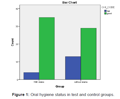
In Group A (Test group), Out of 40 patients with chromogenic black stains, 35 has good oral hygiene, 5 had fair oral hygiene.
In Group B (Control group), Out of 40 patients, 29 had good oral hygiene and 11 had fair oral hygiene [Table 1].
| Variables | OHI_ status | Total | ||
|---|---|---|---|---|
| Fair | Good | |||
| Group | Test | 5 | 35 | 40 |
| Control | 11 | 29 | 40 | |
| Total | 15 | 65 | 80 | |
Table 1: Oral hygiene status.
Chi-square test is statistically significant (p<0.05). There is a significant difference in the oral hygiene scores between groups. The test group (Group A) patients showed better oral hygiene status than the control group (Group B) patients [Table 2 and Figure 1].
| Chi-Square Tests | |||
|---|---|---|---|
| Variables | Value | Df | Asymp. Sig. (2-sided) |
| Pearson Chi-Square | 5.223a | 1 | .022 |
| Continuity Correctionb | 4.050 | 1 | .044 |
| Likelihood Ratio | 5.469 | 1 | .019 |
| Fisher's Exact Test | |||
| N of Valid Cases | 81 | ||
a0 cells (0.0%) have expected count less than 5. The minimum expected count is 8.19.
bComputed only for a 2 × 2 table
Table 2: Comparison of OHIS between the groups.
Plaque index (Silness and Loe)
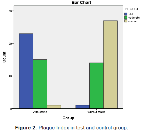
In Group A patients with stains, out of 40 patients, in 24 patients, only a thin film of plaque was detected, 15 patints showed moderate accumulation of plaque, whereas 1 showed abundance of soft matter within the gingival pocket and on tooth surface [Table 3].
| Variables | PI | Total | |||
|---|---|---|---|---|---|
| Mild | Moderate | Severe | |||
| Group | Test | 24 | 15 | 1 | 40 |
| Control | 1 | 13 | 26 | 40 | |
| Total | 25 | 28 | 27 | 80 | |
Table 3: Plaque index scores.
In Group B patients without stains, Out of 40 patients only 1 had a thin film of plaque, 13 had moderate accumulation of plaque whereas 26 showed abundance of soft matter within the gingival pocket and on tooth surface.
Chisquare test shows statistically significant values. A significant difference in the plaque scores were observed between the groups [Table 4].
| Chi-Square Tests | |||
|---|---|---|---|
| Variables | Value | Df | Asymp. Sig. (2-sided) |
| Pearson Chi-Square | 44.294a | 2 | .000 |
| Likelihood Ratio | 55.069 | 2 | .000 |
| N of Valid Cases | 81 | ||
a0 cells (0.0%) have expected count less than 5. The minimum expected count is 11.56.
Table 4: Comparison of plaque index between the groups.
On comparison of plaque indices between the groups, test group (Group A) showed lesser plaque scores compared to control group (Group B) which is statistically significant (p<0.05) [Figure 2].
H/o scaling
In the test group out of 40 patients with black stains, 37 patients gave a history of scaling and polishing, only 3 patients had not undergone any scaling and polishing procedures. 8 patients had undergone the treatment before 1to 2 years, 9 patients before 6 months to 1 year.
18 patients gave a recent history of scaling and polishing (1- 6 months). These findings show that the recurrence rates of these stains are very high. Almost 5 patients showed a recurrence within 1-2 months.
In the control group only 6 patients gave a history scaling and polishing. Out of which 2 patients had undergone the treatment before 3 years, 2 had done before 2 years, one patient had undergone one year before, and one patient before four months [Table 5].
| Chi-Square Tests | |||
|---|---|---|---|
| Variables | Value | df | Asymp. Sig. (2-sided) |
| Pearson Chi-Square | 60.466a | 12 | .000 |
| Likelihood Ratio | 76.058 | 12 | .000 |
| N of Valid Cases | 81 | ||
a24 cells (92.3%) have expected count less than 5. The minimum expected count is .48.
Table 5: Comparison of scaling history between the groups.
Here chi-square test is statistically significant. We observe an association between histories of scaling between the groups. Test group patient (Group A) show frequent history of scaling compared to control group (Group B) p<0.05
Discussion
Black chromogenic stains are a frequent finding in day to day dental practice. It is different in etiology and composition from other types of stains by the presence of insoluble iron salts and high calcium and phosphate composition. The black material is a ferric compound, most likely a ferric sulfide, which arises from the interaction between hydrogen sulfide (produced by the bacteria in the periodontal environment) and iron in the saliva or gingival fluid.
Study by Amirth Teerth et al. found that out of 5 scrapings 3 showed presence of ferrous ions of about 2.56%, calcium ions 17.15% and magnesium ions 0.72%, while the remaining 2 samples showed calcium 14.86%, magnesium ions 0.82% and no presence of ferrous ions. [3]
Surdaka et al. invesitigated on the content of saliva samples determinations were done of sodium, potassium, chlorides, copper, zinc, iron, total calcium, inorganic was found of total calcium, inorganic phosphates, copper, sodium, total protein, and lower glucose content than in controls. [10]
Mesonjesi et al. [11] suggested in iron deficient anemia and in iron overload the concentration of iron present in saliva is much higher than in individuals with no anemia hence extrinsic black stains of teeth may be a sign of iron deficient anemia or iron overload. Anemia has been proved as a risk factor in gingival and periodontal diseases which is associated with frequent gingival bleeding. [12]
Some hypotheses concerning the associations between black stains and some bacterial strains (Actinomyces, Lactobacillussp, prevotella melanogenica) has been reported. [13,14]
Other causes of extrinsic tooth discolouration include smoking, betel nut chewing, use of mouth rinses, intake of iron supplements especially in the form of tonics, use of ayurvedic medicines, intake of tea and coffee. Hence these patients were excluded from the study after a recording a detailed history.
The present study shows that chromogenic black stains are more prevalent in patients with good oral hygiene. Eriksen and Nordobo also confirmed that the black type of staining were normally found in patients with good oral hygiene and can be retentive, particularly around the cervical margins of the teeth.
It sometimes occurs in patients with Mediterranean diets. [15] Hence the findings support the present study.
Dayan et al. reported that poor oral hygiene may result in green, black-brown and orange staining which is produced by chromogenic bacteria. Presence of these stains was also associated with lower plaque scores [16] whereas, the study by Sarkonen et al. and Hataba et al. contradicts the present study. Sarkonen et al. observed that the presence of these stains were more prevalent in females than males and were associated with poor oral hygiene. [5]
A study conducted by Hattaba et al. also reported that Green stain appears as a heavy grey green (almost black), soft and furry film, and has been attributed to fluorescent bacteria and fungi were supposedly caused by chromogenic bacteria and again is found in a population with poor oral hygiene. [6]
These deposits are normally seen in children and are found on the buccal surfaces of maxillary teeth. [15] Interestingly in patients with black stains, the plaque index (Silness and Loe) was lower than the control group. These findings are similar to the study conducted by Tomaz Zyla et al. [17] A study by Xi Chen et al. also reported that the black stains were associated with lower visible plaque index in which the visible plaque index was lower in patients with stains than the control group. [18]
Out of 40 patients with black stains, 37 patients gave a history of scaling and polishing, and only 3 patients had not undergone scaling and polishing. 8 patients had undergone the treatment before 1-2 years, 9 patients before 6 months - 1 year.
18 patients gave a history of scaling and polishing before 1- 6 months. These findings show that the recurrence rates of these stains are very high. Almost 5 patients showed a recurrence within 1-2 months. These findings are in agreement to the study by Yue Li et al. [19,20]
In the control group only 6 patients gave a history of scaling and polishing. These findings suggest that the patients with these black stains incidentally are forced to undergo frequent scaling and polishing at very short intervals, due to their unaesthetic appearance and high rate of recurrence. The inadvertent scaling and polishing might ultimately result in wearing of superficial tooth structure which might prove deleterious if done so frequently (every 1-2 months).
Conclusion
The findings of this study indicate the black chromogenic stains are more prevalent in patients with good oral hygiene and good plaque control. Hence this study shows that plaque and the associated bacteria may not be the sole etiology for these stains. They had a very high recurrence rate and may reappear even one month after their complete removal using scaling and polishing. Hence further studies are required to determine the exact etiology of these stains and appropriate preventive strategies to stop their reccurance.
Conflict of Interest
All authors disclose that there was no conflict of interest.
REFERENCES
- Koch MJ, Bove M, Schroff J, Perlea P, Garcia-Godoy F, Staehle HJ. Black stain and dental caries in school children in Potenza, Italy. ASDCJ Dent Child. 2001;68(5-6):353-5,02.
- Schourie KL. Mesentric line on pigmented plaque: A sign of comparative freedom from caries. J Am Dent Assoc 1947;35:805- 807.
- Tirth A, Srivastava BK, Nagarajappa R, Tangade P, Ravishankar TL. An investigation into black tooth stain among school children in Chakkar Ka Milak of Moradabad City, India. J Oral Health Comm Dent 2009;3(2):34-37.
- Gasperito A, Conrado CA, Maciel SM, Miyamoto EY, Chi-carelli M, et al. (2003) Prevalence of black stains and dental caries in schoolchildren in Brazilian school children. Braz Dent J 14: 157-161.
- Sarkonen N, Kononen E, Summanen P, Kanervo A, Takala A, et al. (2000) Oral colonization with Actinomyces species in infants by two years of age. J Dent Res 79:864-867.
- Hattab FN, Qudeimat MA, Al–Rimawi HS (1999) Dental discoloration: An overview. J Esthet Dent 11:291-310.
- Prathap S, Hegde S, Kashyap R, Prathap MS, Arunkumar MS. Clinical evaluation of porous hydroxyapatite bonegraft (Periobone G) with and without collagen membrane (Periocol) in the treatment of bilateral grade II furcation defects in mandibular first permenant molars; Journal Indian Society of Periodontology 17(2): 228, 2013.
- Lobene RR. Effect of dentrifices on tooth stain with controlled brushing. J Am Dent Assoc 1968; 77:849-855.
- Peter S. Essentials of Public Health dentistry (5th edn).
- Surdacka A. Chemical composition of the saliva in children and adolescents with black tartar;Czas stomatol 2012 79(2):219-21.
- Mesonjesi I. Are extrinsic black stains of teeth iron-saturated bovine lactoferrin and a sign of iron deficient anemia or iron overload? Arch Pediatr. 2011 18(12):1348-52.
- Prathap S, Hegde S, Rajesh KS, Arun MS. Anemia-risk factor for periodontitis. 2010, KDJ 33,173-50.
- Shah HN, Bonnett R, Mateen B, Williams RA (1979) The porphyrin pigmentation of subspecies of Bacteroides melanogeniucus. Biochem J 180 :45-50.
- Siti I, Indiarti Y, Rustan R. Identification of quantitity of Actinomyces in children saliva with black stain in tooth enamel surface. Int J Clin Prev Dent; 2013; 3: 164-168.
- Nordbo HE. Extrinsic discoloration of teeth. J Clin Periodontol 1978, 5:229-36.
- Dayan D, Heifferman A, Gorski M, Beigleiter A (1983) Tooth discolouration: Extrinsic and intrinsic factors. Quintessence Int 12 (14):1-5.
- Zyla T, Kawala B, Antoszewsska J, Smith P, Kawala M. Black stain and dental caries: A review of literature. Biomed Research International Volume 2015. p: 6.
- Chen X, Zhan JY, Lu HX, Ye W, Zhang W, Yang WJ, et al. Factors associated with black tooth stain in Chinese preschool children. Clin Oral Investigations 2014, 9: 2059-2066.
- Li y, Zhang Q, Zhang f, Liu R, Chen F. Analysis of microbiota of black stain in primary dentition. PLoS One 10(9): e0137030.
- Prathap S, Rajesh H, Aoloor V, Rao AS. Extrinsic stains and management: A new insight. J Acad Indus Res; 2013: 435-442.


 The Annals of Medical and Health Sciences Research is a monthly multidisciplinary medical journal.