Paley’s 3 Fibular Hemimelia in a 2 Month Old Infant
Received: 01-Nov-2022, Manuscript No. amhsr-22-82127; Editor assigned: 03-Nov-2022, Pre QC No. amhsr-22-82127 (PQ); Reviewed: 18-Nov-2022 QC No. amhsr-22-82127; Revised: 28-Nov-2022, Manuscript No. amhsr-22-82127 (R); Published: 05-Dec-2022
Citation: Agarwal S. Paleyâ??s 3 Fibular Hemimelia in a 2-Month-Old Infant. Ann Med Health Sci Res. 2022;12:362-363.
This open-access article is distributed under the terms of the Creative Commons Attribution Non-Commercial License (CC BY-NC) (http://creativecommons.org/licenses/by-nc/4.0/), which permits reuse, distribution and reproduction of the article, provided that the original work is properly cited and the reuse is restricted to noncommercial purposes. For commercial reuse, contact reprints@pulsus.com
Case Description
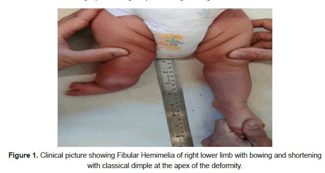
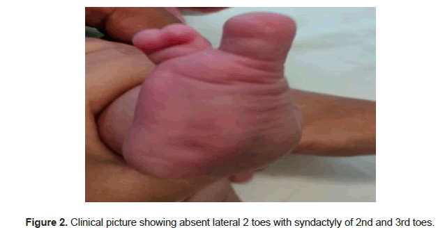
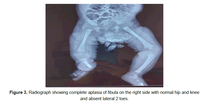
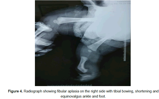
Fibular Hemimelia (FH) is a congenital deficiency where part or all of the fibular bone is hypoplastic, dysplastic or aplastic associated with dysplasia of tibia and parts of the foot [1]. The Paley classification is the first classification of FH to be designed with reconstructive surgery options in mind. In type 3, there is a fixed deformity of equino-valgus. Here, we present a case who was 2 months old. On examination there was anteromedial bowing of tibia with absent fibula and lateral 2 toes and syndactyly of 2nd and 3rd toes. There was shortening of 2 cm, with projected shortening of tibia of 11 cm at maturity based on Paley’s multiplier method (Figures 1-4). Reconstructive surgery is planned in follow up as:
• Surgery#1, at age 18 months, SUPER ankle (Systematic Utilitarian Procedure For Extremity Reconstruction) procedure combined with lengthening of 5.0 cm combined with hemiepiphysiodesis of distal femur for valgus knee correction.
• Surgery #2, at age 8 years, lengthening 6.0 cm of tibia.
References
- Paley D. Surgical reconstruction for fibular hemimelia. J Child Orthop. 2016;10:557-583.
[Crossref] [Google Scholar] [Indexed]


 The Annals of Medical and Health Sciences Research is a monthly multidisciplinary medical journal.