Patients Undergoing Restorative Treatment for Management of Upper Anterior Spacing: A Retrospective Analysis
2 Department of Conservative dentistry and Endodontics, Saveetha Dental College and Hospitals, Saveetha Institute of Medical and Technical Sciences, Saveetha University, Chennai, India, Email: sowmyak.sdc@saveetha.com
3 Department of Pedodontics, Saveetha Dental College and Hospitals, Saveetha Institute of Medical and Technical Sciences, Saveetha University, Chennai, India
Received: 31-Aug-2021 Accepted Date: Sep 13, 2021 ; Published: 20-Sep-2021
This open-access article is distributed under the terms of the Creative Commons Attribution Non-Commercial License (CC BY-NC) (http://creativecommons.org/licenses/by-nc/4.0/), which permits reuse, distribution and reproduction of the article, provided that the original work is properly cited and the reuse is restricted to noncommercial purposes. For commercial reuse, contact reprints@pulsus.com
Abstract
Anterior spacing is a common problem in patients seeking esthetic treatment. The most common etiology for spacing is tooth size and arch length discrepancy. A carefully documented diagnosis and treatment plan are essential if the clinician is to apply the most effective approach to address the patient’s needs. A multidisciplinary approach is sometimes necessary to correct the esthetics and to improve the occlusion. The main aim of this study was to analyse the number of patients choosing restorative treatment for correction of spacing in the upper anterior region over fixed or removable orthodontic treatment. This retrospective study had a sample size of 1613. It was conducted among patients who visited the outpatient Department at the institution with a chief complaint of upper anterior spacing. The data was collected from the patient records. The following data were retrieved from the dental records: patient age, gender, and preference for restorative treatment, fixed or removable orthodontic treatment was analysed. The coding was done in MS excel and data was transferred to a host computer and processed using SPSS software version 20.0 (SPSS Inc., Chicago, IL, USA). Descriptive statistics was used to study the data collected and to analyse frequency distribution. Chi square test was used to assess the association at 5% significance level (P<0.05). The results showed no significant association between either age or gender against the treatment option preferred. Only 2.9% opted for restorative management of upper anterior spacing, 84.6% preferred fixed orthodontic treatment and 12.5% preferred removable orthodontic appliance.
Keywords
Fixed orthodontic appliance; Generalised spacing; Midline diastema; Removable orthodontic appliance; Restorative management
Introduction
Anterior spacing is a major aesthetic concern for most young people today. The most common aetiology for spacing is tooth size and arch length discrepancy. A space between adjacent teeth is called a “diastema”. Midline diastema (or diastases) occurs in approximately 98% of 6 year olds, 49% of 11 year olds and 7% of 12–18 year olds. [1] In most children, the medial erupting path of the maxillary lateral incisors and maxillary canines, as described by Broadbent results in normal closure of this space. [2] In some individuals however, the diastema does not close spontaneously. The continuing presence of a diastema between the maxillary central incisors in adults often is considered an esthetic problem. [3] Maxillary lateral incisors vary more than any other tooth in the dentition. Microdontia is an anomaly where the tooth is abnormally small, when this occurs in the maxillary lateral it is called “peg lateral”. [4] Backman et al., found that peg-shaped maxillary lateral incisors occurred more frequently than other developmental malformations of the teeth, with an incidence of 0.8% in 739 Swedish children. [5] One study of 8,289 students found that 1.78% exhibited either peg-shaping or agenesis of permanent maxillary lateral incisors, with a greater frequency in females. [6]
Spacing in the upper anterior region can be one of the most negative factors in self perceived dental appearance. Treatment is mainly for esthetic and psychological reasons, rather than functional ones. [7] The extent and the etiology of the diastema must be properly evaluated. Proper case selection, appropriate treatment selection, adequate patient cooperation, and good oral hygiene are all important factors for successful treatment outcomes. [8,9] Depending on the etiology, a comprehensive treatment plan needs to be formulated. There are several ways to address this–it can be done through orthodontics or through restorative or interdisciplinary techniques. Restorative measures include laminate veneers, direct veneering, composite build up and full veneer crown.
Orthodontic treatment can be employed when there is a jaw size, arch length and tooth size discrepancy. This provides permanent results if time is not a constraint. However, it requires several follow up appointments and the process takes a longer duration to achieve satisfactory results. [10] Post orthodontic treatment, relapse is a common occurrence if no retention appliance is given, occurrence of whit spot lesions is another possibility, crestal alveolar bone loss and apical root resorption and gingivitis also can occur. [11–14] Due to all the above stated reasons, some patients prefer restorative management to address the spacing.
Among the various restorative options available, porcelain laminate veneers are considered the most conservative, minimally invasive and highly aesthetic approach which has shown superior aesthetics with long term success. [15] Restoring the spacing in anterior teeth can be achieved quickly with direct composite veneering. [16] They are good alternatives to porcelain veneers in teeth with additional surface defects erosion, non-carious lesions [17] or hypoplastic enamel. [18,19] A more invasive treatment like full coverage crowns may require root canal treatment before addressing aesthetic concerns especially in cases with non-vital or decayed teeth. [20–23] Tooth discoloration due to calcific metamorphosis, trauma or endodontic procedures like use of intracanal medicaments or insufficient cleaning and shaping must be carefully evaluated before laminates or full veneer crowns are advised. [22,24,25] Previously our team has a rich experience in working on various research projects across multiple disciplines. [26–40] Now the growing trend in this area motivated us to pursue this project.
It is essential to discuss all options with patients so that they are involved in the decision making process. In simpler cases, either restorative management, fixed or removable orthodontic treatment can be employed independently. Efforts to treat the patient as a whole using a multidisciplinary approach will provide satisfactory results in complicated cases. [41] The aim of this study was to analyse the number of patients choosing restorative treatment for correction of spacing in the upper anterior region over orthodontic treatment.
Materials and Methods
Study setting
In this retrospective study, data from 1613 patients within the department of conservative dentistrywere collected from dental records. At data extraction, all information was anonymized and tabulated onto a spreadsheet. The study was commenced after approval from the institutional review board. The ethical approval number for the study was SDC/SIHEC/2020/DIASDATA/0619-0320.
Data collection and tabulation
To fulfill the inclusion criteria, patients who had upper anterior spacing were included in the study. The preference of treatment options to correct the spacing was assessed in these patients. Patients who did not have spacing and those unwilling for the treatment have been excluded.
Sampling
Data were collected from June 2019 to March 2020 for 1613 patients who reported with upper anterior spacing. The following data were retrieved from the dental records: patient age, gender, and preference for restorative treatment, fixed or removable orthodontic treatment was analyzed.
Statistical analysis
The data was transferred to a host computer and processed using SPSS software version 21.0 (SPSS Inc., Chicago, IL, USA). Descriptive statistics and Chi square test was used to compare the preference for restorative treatment, fixed or removable orthodontic treatment with age and gender of the patient. The significance level was set at 5%.
Results and Discussion
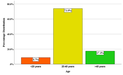
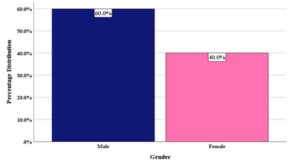
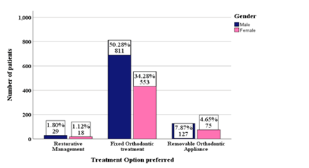
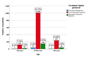
Total number of patients that reported spacing in upper anteriors was 1613. The distribution of age among the study group showed 8.7% of the population below 20 years of age, 73.9% between 20-40 years, and 17.4% above 40 years [Figure 1]. The gender distribution of study participants shows 60% males and 40% females [Figure 2]. The majority of the patients opted for fixed orthodontic treatment 84.6% followed by removable orthodontic appliances 12.5% and the least preferred treatment option was restorative management 2.9% [Figure 3]. No significant association was found between gender and treatment option preferred (P value: 0.633; Chi square test) [Figure 4] or age and treatment option preferred (P value: 0.754; Chi square test) [Figure 5].
Figure 1:Bar diagram representing distribution of study subjects according to age. X-Axis represents the age group distribution and Y axis represents the percentage distribution of different age groups. The percentage distribution shows 8.7% were less than 20 years (orange), 73.9% were between 20-40 years (yellow) and 17.4% were above 40 years (green). Highest number of study subjects reporting with spacing was between 20-40 years (73.9%).
Figure 2:Bar diagram representing distribution of study subjects according to gender. X-Axis represents the gender distribution and Y axis represents the percentage distribution. It shows 60% were males (dark blue) and the remaining 40% (pink) were females. There were more male patients that reported with spacing than female patients.
Figure 3:Bar diagram representing treatment option preferred by patients reporting with spacing. X-Axis represents the treatment option preferred and Y axis represents the percentage distribution in each category. The graph shows 2.9% preferred restorative management (blue), 84.6% preferred fixed orthodontic treatment (red) and 12.5% preferred removable orthodontic treatment (green). The maximum number of individuals preferred fixed orthodontic treatment (84.6%).
Figure 4:Bar diagram representing association between the treatment options preferred and gender of the patients. X-axis represents the preference of treatment option and Y-axis represents the number of patients. Majority of both male (dark blue) and female patients (pink) preferred fixed orthodontic treatments while the least preferred treatment option was restorative treatment among both genders. No significant association was found between the treatment option preferred and the gender of the patients (Pearson’s chi square value 0.916, df-2, p value=0.633 not significant).
Figure 5:Bar diagram representing association between the treatment option preferred and age of the patients. X-axis represents the age of the patient and Y-axis represents the number of patients. Fixed orthodontic treatment (red) was the most preferred treatment among all age groups. The preference for restorative treatment (blue) and removable appliances (green) was highest in the 20-40 year age group. However, no significant association was found between the treatment option preferred and the age of the patients (Pearson’s chi square value 1.903, df-4, p value=0.754 not significant).
Abnormalities in tooth size, shape, and structure result from disturbances during the morpho-differentiation stage of development, and ectopic eruption, hypomineralization, [42] rotation and impaction of teeth result from developmental disturbances or form trauma. [43,44] Morphological abnormalities like peg shaped lateral incisors that contribute to anterior spacing occur more in women than men. [45] However in our study there were more men who reported with spacing than women.
Although very few patients in our study 2.9% opted for restorative management like laminate veneers, direct veneering, composite build up and full veneer crown, it must be noted that restorative management can improve aesthetics when there are developmental anomalies in the tooth itself. Levin used the golden proportion (proportion of 1.618:1.0) in order to achieve an esthetic smile. [46] The low preference rate for restorative management seen in this study [Figure 3] could be due to lack of awareness on the aesthetic outcomes, longevity of the veneers and the cost factor associated with it. Introduction of special acid etching techniques and advancements in the bonding system has improved the long-term retention and survival rates for veneers and laminates. [47] Porcelain laminate veneers are more esthetic than direct or indirect composite veneers and are also considered to be more conservative. Only a small amount of enamel reduction on the labial surface is needed to create a definitive 0.5 mm margin and surface roughness. Maintaining adequate biological width prevents gingival inflammation and subsequent damage to periodontium. [48] Direct composite veneers have shown a higher risk of failure compared with porcelain veneers at a 2.5 year evaluation. [49] Another study by Peumans and colleagues revealed an excellent retention rate of porcelain laminate veneers after 10 years, with only 4% of 87 veneers having to be replaced at follow-up. [50] Dental plaster models and dental photographs allow dentists to examine and study the occlusion proportions of the teeth. A diagnostic wax-up can display the desired treatment outcome and thus can be visualized by both the practitioner and the patient. The esthetic result of ceramic veneers was good when maintained well, [51,52] with high patient satisfaction. [53]
In our study, 84.6% of the study population preferred fixed orthodontic treatment followed by 12.5% who preferred a removable appliance for the management of their spacing [Figure 3]. This could be due to the fact that orthodontics offers versatile treatment modalities for management of spacing. Also, patients of younger age groups may find the long duration of orthodontic treatment acceptable and the preference for orthodontic treatment could be attributed to the majority of patients being less than 40 years of age in this study [Figure 1 and Figure 5]. Number of male patients with upper anterior spacing was more than females in this study, but no significant association was found in the treatment option preferred among males and females [Figure 2 and Figure 4].
Nonetheless, an orthodontic treatment can only align the teeth in their respective position in the arch and fails to address anomalies like peg laterals or congenitally missing teeth. However when lip profile, proclination and arch need to be corrected then restorative management will not yield satisfactory results and orthodontics come into play. Fixed orthodontic treatment can cause bodily movement of the tooth, translation, intrusion, extrusion and allows a more precise tooth movement while removable orthodontic appliances are suitable for minor tooth movement using tipping force applied to the individual tooth. [54] Esthetics as well as occlusion must be considered during treatment planning. [55] Our institution is passionate about high quality evidence based research and has excelled in various fields. [56-61] We hope this study adds to this rich legacy. Treatment in esthetic cases often involves a multidisciplinary approach, such as orthodontic treatment, periodontal evaluation, oral surgery, restorative treatment, and prosthodontics. To achieve the desired esthetically pleasing treatment, smile analysis is essential and golden proportions must be followed to achieve optimal results. Small sample size, geographic isolation, no recording of family history, type of spacing (midline diastema, peg laterals, congenitally missing teeth, trauma etc.) and lack of inclusion of socio economic factors are the limitations of this study which can be focused in future studies.
Conclusion
Within the limitations of the study, it can be concluded that age and gender had no influence on the treatment option preferred for anterior spacing. Most patients prefer orthodontic treatment over restorative treatment for treating anterior spacing. The restorative treatment, whenever considered, must preserve as much of the original tooth structure as possible. Time is a deciding factor for preference in choice of treatment. Patients with anterior spacing must be carefully evaluated for interdisciplinary treatment planning to obtain excellent results.
Author Contributions
Author 1 (Jerusha Santa Packyanathan) carried out the retrospective study by collecting data and drafted the manuscript after performing the necessary statistical analysis. Author 2 (Sowmya K) aided in the conception of the topic, participated in the study design, statistical analysis, supervised in the preparation of the manuscript and author 3 (Ganesh Jeevanandan) helped in study design and coordinated in developing the manuscript. All the authors have equally contributed in developing the manuscript.
Acknowledgement
The authors would like to acknowledge the support rendered by the department of conservative dentistry and endodontics and Information and technology department of Saveetha dental college and hospitals and the management for their constant assistance with the research.
REFERENCES
- Foster TD, Grundy MC. Occlusal changes from primary to permanent dentitions. Br J Orthod. 1986;13:187-193.
- Broadbent BH. The face of the normal child. Angle Orthod. 1937;7:183-208.
- Ferguson MW, Rix C. Pathogenesis of abnormal midline spacing of human central incisors. A histological study of the involvement of the labial frenum. Br Dent J. 1983;154:212-218.
- Shrestha BK, Yadav R, Gupta S. Prevalence of malocclusion among medical students in Institute of Medicine, Nepal: A preliminary report. Orthod J Nepal. 2011;1.
- Bäckman B, Wahlin YB. Variations in number and morphology of permanent teeth in 7-year-old Swedish children. Int J Paediatr Dent. 2001;11:11-17.
- Meskin LH, Gorlin RJ. Agenesis and peg-shaped permanent maxillary lateral incisors. J Dent Res. 1963;42:1476–1479.
- Bernabe E, Flores-Mir C. Influence of anterior occlusal characteristics on self-perceived dental appearance in young adults. Angle Orthod. 2007;77:831–836.
- Abrahams R, Kamath G. Midline diastema and its aetiology–A review. Dent Update. 2014;41:457–464.
- Bishara SE. Management of diastemas in orthodontics. Am J Orthod. 1972;61:55–63.
- Chan MD. An adult malocclusion requiring a combination of orthodontic and prosthodontic treatment. Am J Orthod Dentofacial Orthop. 1997;111:100–105.
- Levin L, Samorodnitzky‐Naveh GR. The association of orthodontic treatment and fixed retainers with gingival health. J Periodontol. 2008;79:2087-92.
- Bergstrand F, Twetman S. A review on prevention and treatment of post-orthodontic white spot lesions–evidence-based methods and emerging technologies. Open Dent J. 2011;5:158-162.
- Sharpe W, Reed B, Daniel Subtelny J. Orthodontic relapse, apical root resorption, and crestal alveolar bone levels. Am J Orthod Dentofacial Orthop. 1987;91:252–258.
- Zawawi KH, Melis M. The role of mandibular third molars on lower anterior teeth crowding and relapse after orthodontic treatment: A systematic review. Sci World J. 2014.
- Ravinthar K. Recent advancements in laminates and veneers in dentistry. Research J Pharm and Tech. 2018;11:785-787.
- Garber D. Porcelain Laminate Veneers: Ten Years Later Part I: Tooth Preparation. J Esthet Dent. 1993;5:57–62.
- Nasim I, Hussainy S, Thomas T, et al. Clinical performance of resin-modified glass ionomer cement, flowable composite, and polyacid-modified resin composite in noncarious cervical lesions: One-year follow-up. J Conserv Dent. 2018;21:510-515.
- Mahalakshmi N, Nasim I. Comparative evaluation of grape seed and cranberry extracts in preventing enamel erosion: An optical emission spectrometric analysis. J Conserv Dent. 2018;21(5):516–520.
- Jose J, Subbaiyan H. Different Treatment Modalities followed by Dental Practitioners for Ellis Class 2 Fracture–A Questionnaire-based Survey. Open Dent J. 2020;14:59-65.
- Ramanathan S, Solete P. Cone-beam computed tomography evaluation of root canal preparation using various rotary instruments: An in vitro Study. J Contemp Dent Pract. 2015;16:869-872.
- Ramamoorthi S, Nivedhitha MS, Divyanand MJ. Comparative evaluation of postoperative pain after using endodontic needle and endoactivator during root canal irrigation: A randomised controlled trial. Aust Endod J. 2015;41:78–87.
- Manohar MP, Sharma S. A survey of the knowledge, attitude, and awareness about the principal choice of intracanal medicaments among the general dental practitioners and nonendodontic specialists. Indian J Dent Res. 2018;29:716-720.
- Janani K, Palanivelu A, Sandhya R. Diagnostic accuracy of dental pulse oximeter with customized sensor holder, thermal test and electric pulp test for the evaluation of pulp vitality: an in vivo study. Braz Dent. 2020;23:8.
- Kumar D, Delphine Priscilla Antony S. Calcified Canal and Negotiation-A Review. Research Journal of Pharmacy and Technology. 2018;11:3727-3730.
- Teja KV, Ramesh S. Shape optimal and clean more. Saudi Endod J. 2019;9:235-236.
- Ponnulakshmi R, Shyamaladevi B, Vijayalakshmi P, Selvaraj J. In silico and in vivo analysis to identify the antidiabetic activity of beta sitosterol in adipose tissue of high fat diet and sucrose induced type-2 diabetic experimental rats. Toxicol Mech Methods. 2019;29:276–290.
- Mathew MG, Samuel SR, Soni AJ, Roopa KB. Evaluation of adhesion of Streptococcus mutans, plaque accumulation on zirconia and stainless steel crowns, and surrounding gingival inflammation in primary molars: randomized controlled trial. Clin Oral Investig. 2020;24:3275–3280.
- Subramaniam N, Muthukrishnan A. Oral mucositis and microbial colonization in oral cancer patients undergoing radiotherapy and chemotherapy: A prospective analysis in a tertiary care dental hospital. J Investig Clin Dent. 2019;10:e12454.
- Girija ASS, Shankar EM, Larsson M. Could SARS-CoV-2-Induced hyperinflammation magnify the severity of coronavirus diseae(CoViD-19) leading to acute respiratory distress syndrome? Front Immunol. 2020;11:1206.
- Dinesh S, Kumaran P, Mohanamurugan S, et al. Influence of wood dust fillers on the mechanical, thermal, water absorption and biodegradation characteristics of jute fiber epoxy composites. J Polym Res. 2020;27.
- Thanikodi S, Singaravelu D Kumar, Devarajan C, Vijayan V, Venkatesh R. Teaching learning optimization and neural network for the effective prediction of heat transfer rates in tube heat exchangers. Therm Sci. 2020;24:575–581.
- Murugan MA, Jayaseelan V, Jayabalakrishnan D, Maridurai T, Kumar SS, Ramesh G et al. Low velocity impact and mechanical behaviour of shot blasted SiC wire-mesh and silane-treated aloevera/hemp/flax-reinforced SiC whisker modified epoxy resin composites. Silicon Chem. 2020;12:1847–1856.
- Vadivel JK, Govindarajan M, Somasundaram E, et al. Mast cell expression in oral lichen planus: A systematic review. J Investig Clin Dent. 2019;10:e12457.
- Chen F, Tang Y, Sun Y, Veeraraghavan VP, Mohan SK, Cui C. 6-shogaol, a active constiuents of ginger prevents UVB radiation mediated inflammation and oxidative stress through modulating NrF2 signaling in human epidermal keratinocytes (HaCaT cells). J Photochem Photobiol B. 2019;197: 111518.
- Manickam A, Devarasan E, Manogaran G, Priyan MK. Score level based latent fingerprint enhancement and matching using SIFT feature. Multimed Tools Appl. 2019;78:3065–3085.
- Wu F, Zhu J, Li G, Wang J, Veeraraghavan VP, Mohan SK, et al. Biologically synthesized green gold nanoparticles from induce growth-inhibitory effect on melanoma cells (B16). Artif Cells Nanomed Biotechnol. 2019;47:3297–3305.
- Ma Y, Karunakaran T, Veeraraghavan VP, Mohan SK, Li S. Sesame inhibits cell proliferation and induces apoptosis through inhibition of STAT-3 translocation in thyroid cancer cell lines (FTC-133). Biotechnol Bioprocess Eng. 2019;24:646–652.
- Ponnanikajamideen M, Rajeshkumar S, Vanaja M, Annadurai G. In vivo type 2 diabetes and wound-healing effects of antioxidant gold nanoparticles synthesized using the insulin plant chamaecostus cuspidatus in albino rats. Can J Diabetes. 2019;43:82–89.
- Vairavel M, Devaraj E, Shanmugam R. An eco-friendly synthesis of Enterococcus sp.-mediated gold nanoparticle induces cytotoxicity in human colorectal cancer cells. Environ Sci Pollut Res Int. 2020;27:8166–8175.
- Paramasivam A, VijayashreePriyadharsini J, Raghunandhakumar S. N6-adenosine methylation (m6A): a promising new molecular target in hypertension and cardiovascular diseases. Hypertens Res. 2020;43:153–154.
- Nestel E, Walsh JS. Substitution of a transposed premolar for a congenitally absent lateral incisor. Am J Orthod Dentofacial Orthop. 1988;93:395–399.
- Rajendran R, Kunjusankaran RN, Sandhya R. Comparative evaluation of remineralizing potential of a paste containing bioactive glass and a topical cream containing casein phosphopeptide-amorphous calcium phosphate: An in vitro Study. Pesquisa Brasileiraem Odontopediatria e Clínica Integrada. 2019;19:1–10.
- Tai K, Park JH, Takayama A. Congenitally Missing Maxillary Lateral Incisor Treated with Atypical Extraction Pattern. J Clin Pediatr Dent. 2011;36:11–18.
- Rajakeerthi R, Ms N. Natural product as the storage medium for an avulsed tooth–A systematic review. Cumhuriyet Dent J. 2019;22:249-256.
- Gupta SK, Saxena P, Jain S, Jain D. Prevalence and distribution of selected developmental dental anomalies in an Indian population. J Oral Sci. 2011;53:231–238.
- Levin EI. Dental esthetics and the golden proportion. J Prosthet Dent.1978;40:244–252.
- Kihn PW, Barnes DM. The clinical longevity of porcelain veneers: A 48-month clinical evaluation. J Am Dent Assoc. 1998;129:747–752.
- Teja KV, Ramesh S, Priya V. Regulation of matrix metalloproteinase-3 gene expression in inflammation: A molecular study. J Conserv Dent. 2018;21:592-596.
- Meijering AC, Creugers NHJ, Roeters FJM, Miulder J. Survival of three types of veneer restorations in a clinical trial: a 2.5-year interim evaluation. J Dent. 1998;26:563–568.
- Peumans M, De Munck J, Fieuws S, Lambrechts P, Vanherle G, Van Meerbeek B. A prospective ten-year clinical trial of porcelain veneers. J Adhes Dent. 2004;6:65-76.
- Siddique R, Sureshbabu NM, Somasundaram J, Jacob B, Selvam D. Qualitative and quantitative analysis of precipitate formation following interaction of chlorhexidine with sodium hypochlorite, neem, and tulsi. J Conserv Dent. 2019;22:40-47.
- Noor S. Chlorhexidine: Its properties and effects. Res J Pharm Technol. 2016;9:1755–1760.
- Ittipuriphat I, Leevailoj C. Anterior space management: interdisciplinary concepts. J Esthet Restor Dent. 2013;25:16–30.
- Singh G. Textbook of Orthodontics. Jaypee Brothers, Medical Publishers Pvt. Limited. 2008.
- Gupta SP. Management of Anterior Spacing with Peg Lateral by Interdisciplinary Approach: A Case Report. Orthod J Nepal 2019;9:67–73.
- Vijayashree PJ. In silico validation of the non-antibiotic drugs acetaminophen and ibuprofen as antibacterial agents against red complex pathogens. J Periodontol. 2019;90:1441–1448.
- Ezhilarasan D, Apoorva VS, Ashok VN. Syzygiumcumini extract induced reactive oxygen species-mediated apoptosis in human oral squamous carcinoma cells. J Oral Pathol Med. 2019;48:115–121.
- Ramesh A, Varghese S, Jayakumar ND, Malaiappan S. Comparative estimation of sulfiredoxin levels between chronic periodontitis and healthy patients-A case-control study. J Periodontol. 2018;89:1241–1248.
- Sridharan G, Ramani P, Patankar S, Vijayaraghavan R. Evaluation of salivary metabolomics in oral leukoplakia and oral squamous cell carcinoma. J Oral Pathol Med. 2019;48:299–306.
- Pc J, Marimuthu T, Devadoss P. Prevalence and measurement of anterior loop of the mandibular canal using CBCT: A cross sectional study. Clin Implant Dent Relat Res. 2018;20:531-534.
- Ramadurai N, Gurunathan D, Samuel AV, Subramanian E, Rodrigues SJL. Effectiveness of 2% Articaine as an anesthetic agent in children: randomized controlled trial. Clin Oral Investig 2019;23:3543–3550.


 The Annals of Medical and Health Sciences Research is a monthly multidisciplinary medical journal.