Pattern of Fibro-osseous Lesions of Jaws in Port Harcourt in South-South Nigeria
2 Department of Pathology, Bayero University Kano, Aminu Kano Teaching Hospital, Kano, Nigeria
Citation: Iyogun CA, et al. Pattern of Fibro-osseous Lesions of Jaws in Port Harcourt: South-South Nigeria. Ann Med Health Sci Res. 2018;8:186-188
This open-access article is distributed under the terms of the Creative Commons Attribution Non-Commercial License (CC BY-NC) (http://creativecommons.org/licenses/by-nc/4.0/), which permits reuse, distribution and reproduction of the article, provided that the original work is properly cited and the reuse is restricted to noncommercial purposes. For commercial reuse, contact reprints@pulsus.com
Abstract
Background: Fibro-osseous lesions comprise a group of disorders characterized histologically by replacement of normal bone by cellular fibrous tissue within which varying amount of predominantly woven bone and acellular islands of mineralized tissue develops. These lesions commonly affect the jaw and craniofacial bones. Identification of the specific entity is crucial due to treatment variations. This study aimed to describe the spectrum, age, sex and morphological pattern of fibro-osseous lesions in Port Harcourt, as well as compare the findings with previous studies done in Nigeria and other parts of the world. Materials and Methods: This was a 7 year retrospective study from 2nd January, 2008 to 31st December, 2014 of all fibro-osseous lesions recorded in the oral pathology/pathology registers of the University of Port Harcourt Teaching Hospital, Port Harcourt, Nigeria. Results: Thirty one cases of fibro-osseous lesions were diagnosed during the seven year study period, with age range from 6–65 years and mean age of 26.7 years. Female to male ratio was 1.8 : 1. Lesions were more frequent in the mandible (51.6%), than maxilla (45.2%) and frontal bone (3.2%). Ossifying fibroma accounted for the vast majority (21 cases, 67.7%) of fibro-osseous lesions distantly followed by fibrous dysplasia (6 cases, 19.4%) and other less frequent types. Conclusion: This study showed that ossifying fibroma was the predominant subtype which was consistent with most published reports in developing world. Due to close architectural structures of these lesions, a definitive diagnosis requires a thorough knowledge and correlation of clinical, radiological and histological findings.
Keywords
Fibro-osseous lesions; Ossifying fibroma; Fibrous dysplasiaIntroduction
Fibro-osseous lesions comprise a group of disorders characterized histologically by replacement of normal bone by cellular fibrous tissue within which varying amount of predominantly woven bone and acellular islands of mineralized tissue develops. [1] These lesions commonly affect the jaw and craniofacial bones. The histopathology of all fibro-osseous lesions are identical, although they range widely in clinical behavior. [2]
This group of lesions include ossifying fibroma, fibrous dysplasia, osseous dysplasia and other infrequent subtypes. [3] Ossifying fibroma is a fibro-osseous lesion which can occur in any facial bone and it usually affects patients in their third and fourth decade and when it occur in the young, it is termed juvenile ossifying fibroma. It can be distinguished from fibrous dysplasia in that it is usually well demarcated. [4] Fibrous dysplasia on the other hand is usually first recorgnised in the second decade. It could be monostotic, craniofacial or polyostotic when it involved numerous bones. [5]
There is paucity of reports on fibro-osseous lesions in our setting. We therefore undertook this review to document and evaluate the pattern in Port Harcourt, South-South geopolitical zone of Nigeria, as well as to compare the findings with previous studies done in Nigeria and other geographical locations of the world.
Material and Methods
This was a 7-year retrospective study from 2nd January, 2008 to 31st December, 2014 of all fibro-osseous lesions recorded in the Oral Pathology of the University of Port Harcourt/ Teaching Hospital registers of the University of Port Harcourt Teaching Hospital, Port Harcourt, Nigeria. The following variables were obtained; age, sex, frequency and histopathological diagnosis.
Histopathology slides on all cases were retrieved and reviewed by the study authors. Fresh sections were cut from archival paraffin blocks when slides could not be retrieved. All specimens had been fixed in 10% formal saline, routinely processed and analysed. Diagnosis was based on world health organization new classification of fibro-osseous lesions. [6] The data was subsequently analysed using SPSS version 20 and presented as frequency tables.
Results
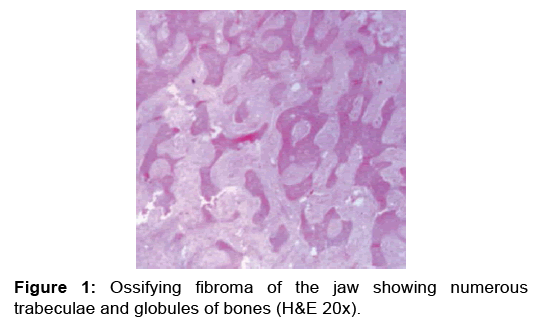
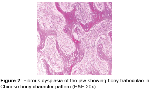
Thirty one cases of fibro-osseous lesions were diagnosed during the seven year study period, with age range from 6–65 years, mean age of 26.7 years and peak age of incidence occurred in the 21–30 years group with 10 cases (32%) and closely followed by 11–20 years with 9 cases (29%). The youngest incidence was a 6 year old. There was slight female preponderance with an overall F: M ratio of 1.8: 1. Tables 1 and 2 depict the relative frequency, age distribution of different histopathologic subtypes and sex distribution of fibo-osseous lesions in Port Harcourt respectively. Lesions were more frequent in the mandible (51.6%), than maxilla (45.2%) and frontal bone (3.2%). Ossifying fibroma accounted for the vast majority [21 cases, 67.7%, Figure 1] of fibro-osseous lesions distantly followed by fibrous dysplasia [6 cases, 19.4%, Figure 2] and other less frequent types. Table 3 showed site distribution of fibo-osseous lesions in the facial bones of 31 patients.
| Fibro-osseous lesions | 1 – 10 years | 11 - 20 years | 21 – 30 years | 31 – 40 years | 41 – 50 years | 51 – 60 years | 61 – 70 years | N (Freq in %) |
|---|---|---|---|---|---|---|---|---|
| Ossifying fibroma | - | 7 | 7 | 3 | 2 | - | 2 | 21 (67.7) |
| Fibrous dysplasia | 2 | 2 | 1 | - | 1 | - | - | 6 (19.4) |
| Osseous dysplasia | 2 | - | 2 | - | - | - | - | 4 (12.9) |
| Total | 4 | 9 | 10 | 3 | 3 | - | 2 | 31 (100) |
Table 1: Relative frequency and age distribution of fiboosseous lesions in Port Harcourt.
| Fibro-osseous lesions | Male | Female | N (Frequency in %) |
|---|---|---|---|
| Ossifying fibroma | 6 | 15 | 21 (67.7) |
| Fibrous dysplasia | 4 | 2 | 6 (19.4) |
| Osseous dysplasia | 1 | 3 | 4 (12.9) |
| Total | 11 | 20 | 31 (100) |
Table 2: Sex distribution of fibo-osseous lesions in Port Harcourt.
| Fibro-osseous lesions | Mandible | Maxilla | Frontal bone | N (Freq in %) |
|---|---|---|---|---|
| Ossifying fibroma | 11 | 10 | - | 21 (67.7) |
| Fibrous dysplasia | 4 | 2 | - | 6 (19.4) |
| Osseous dysplasia | 1 | 2 | 1 | 4 (12.9) |
| Total | 16 | 14 | 1 | 31 (100) |
Table 3: Site distribution of fibo-osseous lesions in the facial bones of 31 patients.
Discussion
A total of thirty one cases of fibro-osseous lesions were seen during the study period, in which ossifying fibroma emerged the most overwhelming preponderant comprising 67.7% of all fibro-osseous lesions. This corroborates most Nigerian studies, 50.4% in Ibadan and 68.3% in Kano. [7,8] In Ghana it constituted 61.5%. [9] This finding is at variance with reports from western communities where fibrous dysplasia accounted for 67% of all fibro-osseous lesions. [10] The reason for this variation in incidence is not very clear.
Ossifying fibromas are considered as benign fibro-osseous neoplasms which are principally encountered in the jaw bones. Although the cell of origin of ossifying fibroma is unknown, they may be derived from elements contained within the periodontal ligament space. Genetic aberrations are being linked to fibro-osseous lesions and that of ossifying fibroma is said to be HPRT2. [11] The radiologic appearance of most ossifying fibromas varies with the stage of development and the amount of bone matrix within the lesion. It usually appears as unilocular mixed radiolucent and radiopaque lesions with sharply defined boarders. [12] Histologically, ossifying fibroma showed prominent calcified structures (ossicles and cementicles) that appeared as eosinophillic or basophillic spherules of bone within a moderately cellular, dense stroma. [13] In this series, ossifying fibroma were seen most predominantly between the 11-20 and 21-30 years age categories, this agrees with most reports, but others have shown that it can occur at any age. [10,14] The appraisal showed female predominance with a female to male ratio of 2.5:1 which is in keeping with other reports. [15] There was almost equal site predilections with 11 and 10 cases occurring in mandible and maxilla respectively, this is at variance with most reports where mandible is the prefer site. [12,16]
Fibrous dysplasia is an anomaly of bone development characterized by harmatomatous proliferation of fibrous tissue within the medullary bone with secondary bone metaplasia, producing immature, newly formed and weakly calcified bone. It is a benign bone disorder of an unknown etiology, uncertain pathogenesis and diverse histopathology. [5] Radiologically, fibrous dysplasia shows radio-dense opacities with a ground glass appearance that blends into surrounding normal bone. [3] Histologically, it is characterised by a stroma of fibroblasts producing bony matrix without evidence of osteoblastic cells at periphery of bony spicules (osteoblastic rimming). This arrangement is sometimes described as Chinese letter pattern. [13] In this review, the peak age groups of fibrous dysplasia were 1–10 and 11–20 years which approximate the span from separate studies in other developing countries and elsewhere. [14,17] While the male predominance displayed in this study with male to female ratio of 2 : 1 correlate other reports, others documented female preponderance. [16,18,19] Mandible was the most common location which is at variance with most other reports. [10,20]
In this appraisal, there were only four cases of osseous dysplasia which may be due to their infrequent occurrence. [10] Our study was not free from the limitations of retrospective institutionalbased studies. This prevalence is likely a tip of iceberg of the true prevalence because not all patients avail themselves to orthodox medical care in our environment. This results in under-estimation of these lesions in our locality. Likewise, not all tissues get to our institution by virtue of the vastness of the study domain and attached cost histology incurred.
Conclusion
This study showed in conclusion, that ossifying fibroma was the most frequently occurring subtype of fibro osseous lesions of the jaws. The epidemiological variables remain similar to what obtains elsewhere in the developing countries. The close architectural structures of these lesions signal the needs for more advanced imaging, molecular testing and immunological assessment in making accurate diagnosis of these lesions
Conflict of Interest
I declare there is no conflict of interest.
REFERENCES
- Brannon RB, Fowler CB. Benign fibro-osseous lesions: A review of current concepts. Adv Anat Pathol. 2001;8:126-143.
- Hall G. Fibro-osseous lesions of head and neck. Diagnostic Histopathology. 2012;18:149-158.
- MacDonald-Jankowski DS. Fibro-osseous lesions of the face and jaws. Clin Radiol. 2004;59:11-25.
- Barclay R. Lucas pathology of tumours of oral tissue. Fibrous dysplasia of bone and ossifying fibroma. 4th edn. Churchill Livingstone, London, UK: 1984;395-340.
- Greco MA, Steiner GC. Ultrastructure of fibrous dysplasia of bone: a study of its fibrous, osseous, and cartilaginous components. Ultrastruct Pathol 1986;10:55-66.
- Rajpal K, Agarwal R, Chhabra R, Bhattacharya M. Updated classification schemes for Fibro-osseous lesions of the oral & maxillofacial region: A review. IOSR Journal of Dental and Medical Sciences. 2014;13:99-103.
- Lasisi TJ, Adisa AO, Olusanya AA. Fibro-osseous lesions of the jaws in Ibadan, Nigeria. Oral Health Dental Manag. 2014;13:41-44.
- Sule AA, Iyogun CA, Adeyemi TE. Pattern of Fibro-osseous Lesions of the Jaws in Kano, Northern Nigeria. J Dent Oral Health 2017;3:1-3.
- Abdulai AE, Gyasi RK. Iddarissu MI. Benign fibroosseous lesions of the facial skeleton: Analysis of 52 cases seen at the Korle Bu Teaching Hospital. Ghana Medical J. 2004;38:96-100.
- Eversole LR. Craniofacial fibrous dysplasia and ossifying fibroma. Oral Maxillofac Surg Clin North Am. 1997;9:625-642.
- Pimenta FJ, Silveira LF, Tavares GC, Silva AC, Perdigão PF, Castro WH, et al. HRPT2 gene alterations in ossifying fibroma of Tavares GC, the jaws. Oral Oncol 2006;42:735–759.
- Jayachandran S, Sachdeva S. Cemento ossifying fibroma of mandible: Report of two cases. J Indian Acad Oral Med Radiol 2010;22:53-56.
- Voytek TM, Ro JY, Edeiken J, Ayala AG. Fibrous dysplasia and cemento-ossifying fibroma. A histologic spectrum. Am J Surg Pathol 1995;19:775-781.
- Langdon JD, Rapidis AD, Patel MF. Ossifying fibroma - One disease or six? An analysis of 39 fibro-osseous lesions of the jaws. Br J Oral Surg.1976;14:1-11.
- Waldron CA. Fibro-osseous lesions of the jaws. J Oral Maxillofac Surg. 1993;51:828-835.
- Adekeye EO, Edwards MB, Goubran GF. Fibro-osseous lesions of the skull, face and jaws in Kaduna, Nigeria. Brit J Oral Surg. 1980;18:57-72.
- Prabhu S, Sharanya S. Naik PM. Fibro-osseous lesions of oral and maxilla-facial region: Retrospective analysis for 20 years. J Oral Maxillofac Pathol. 2013;17:36-40.
- Ajagbe HA, Daramola JO. Fibro-osseous lesions of the jaw: A review of 133 cases from Nigeria. J Natl Med Assoc. 1983;75:593-598.
- Dahlgren SE, Lind PO, Lindbom A, Martensson G. Fibrous dysplasia of the jaw bones. A clinical, Roentgenographic and histological study. Acta Otolaryngology. 1969;68:257-270.
- Bustamante EV, Albiol GL, Aytes BL, Escoda CG. Benign fibro-osseous lesions of the maxilla: Analysis of 11 cases. Med Oral Patol Oral Cir Bucal. 2008;13:653-656.


 The Annals of Medical and Health Sciences Research is a monthly multidisciplinary medical journal.