Perceptive Role of Ultrasonography in Diagnostic Challenge Cases: A Case Report
Citation: Reddy KVK, et al. Perceptive Role of Ultrasonography in Diagnostic Challenge Cases: A Case Report. Ann Med Health Sci Res. 2018; 8: 16-18
This open-access article is distributed under the terms of the Creative Commons Attribution Non-Commercial License (CC BY-NC) (http://creativecommons.org/licenses/by-nc/4.0/), which permits reuse, distribution and reproduction of the article, provided that the original work is properly cited and the reuse is restricted to noncommercial purposes. For commercial reuse, contact reprints@pulsus.com
Abstract
Muscular hypertrophy is a rare benign soft tissue condition of unknown etiology, whereas Lipomas are common benign soft tissue lesions occurring about 15-20% in the head and neck region. Clinically both appear soft, smooth and painless and frequently encountered at middle third of the face. Here, we report an unusual case of benign tumor in a 16-year old boy.
Keywords
Benign tumor; Lipoma; Muscular hypertrophy
Introduction
Muscular hypertrophy is a rare benign condition of idiopathic cause, in the head and neck region. The most commonly involved muscle is Masseter followed by Temporalis and Pterygoid muscles. [1] In 1880, Legg was the first person to describe a case of idiopathic Masseteric hypertrophy and reported on a 10-year old boy. [2] Masseteric hypertrophy is congenital or acquired in origin. [3] Clinically, unilateral or bilateral asymptomatic enlargement of the Masseter muscle is seen with highest incidence in second or third decade of life, without any gender predilection. [4] The unilateral representation results that the patients chew or clench frequently on one side, and other various factors such as malocclusion, bruxism and temporomandibular joint disorders influences this condition. [1,4] Lipomas are the most common soft tissue benign mesenchymal neoplasms accounting 15-20% in the head and neck region. Clinically, represented as slowly growing painless lesion with a soft, sessile or pedenculated and smooth-surfaced with yellow discoloration. [5] The most common site of occurrence is the areas of fat accumulation such as cheek, buccal mucosa and lips. [5]
Case History
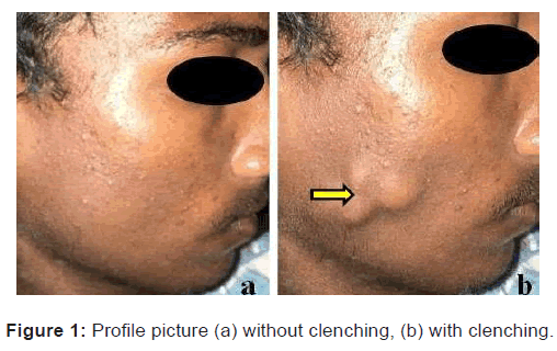
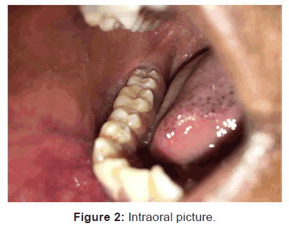
A 16-year-old male patient reported to the department of Oral Medicine and Radiology, with a complaint of unilateral representation of swelling was more evident on clenching on the right side of the face since one year. He had incidentally noticed the swelling was more evident on clenching which remained of the same size since six months, after which it became more bulging. The swelling is not associated with pain and gives no history of trauma. There is no history of any such swellings in other parts of the body. Medical and family history was non-contributory. Clinical examination revealed a well-defined lobulated swelling on the right side of the face measuring about 4 × 3 cm in dimension, which was non-tender and soft in consistency. The swelling was more evident on clenching teeth, no local rise of temperature and mobile in the horizontal direction. Overlying skin was normal, pinchable with no discharge and surrounding skin is normal [Figures 1a and 1b]. Intraoral examination revealed no abnormality [Figure 2]. Based on patient’s history and clinical examination a provisional diagnosis of Masseteric hypertrophy was made with a differential diagnosis of buccal lymphadenitis, soft tissue abscess, Lipoma, accessory parotid tumor and lymphovascular tumor.
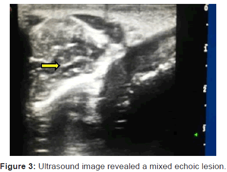
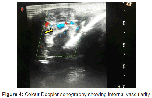
The necessary baseline investigations were done which were non-contributory. Orthopantomograph findings were nonsignificant, ultrasonography showed a 2.2 × 0.6 cm mixed echoic lesion within the right side of masseter muscle [Figure 3]. Colour Doppler ultrasound showed dilated vascular channels with good ?ow [Figure 4], suggestive of venous hemangioma. MRI (magnetic resonance imaging) was not performed due to financial status of the patient; as well he was not willing.
Considering the age of the patient treatment was not attempted. Patient was further followed up for one year to evaluate the status of the lesion which was stable with no further progression.
Discussion
Hemangiomas are rare vascular neoplasms accounting 7% of all benign tumors. [6] These are considered to be tumors of infancy that are characterized by a rapid growth phase with endothelial cell proliferation followed by gradual involution. [5] 60% of the hemangiomas occur in the head and neck region, in which its occurrence in the skeletal muscles is very rare accounting to 0.8% of all hemangiomas. [7,8] The masseter muscle is the most commonly involved and accounting for 5% of all intramuscular hemangiomas followed by trapezius, extraocular muscle, sternocleiodmastoid and temporalis muscle respectively. [9] In 1843, Listen was the first person to report a case of Masseteric intramuscular hemangioma. [10] The exact etiology remains unknown, but according to few others it is a congenital origin due to abnormal embryonic sequestrations or trauma play a role. [11] Intramuscular hemangiomas develop most commonly in the second decade of life and predominantly in males. [12] In 98% of the cases, clinically they represent a firm mass or palpable fluctuant swelling with or without pain. [13] The present case report is consistent with the literature.
Most of the hemangiomas can be diagnosed on clinical examination and not required any imaging modalities unless it is deep seated or clinically atypical soft tissues masses. Conventional radiographs are used as a screening tool for these types of lesions. Ultrasonography (Colour Doppler) and MRI (magnetic resonance imaging) are most frequently used for soft tissue lesions. In the present case conventional radiographs like OPG revealed no evident of any calcifications, ultrasonography revealed a mixed echo lesion and on colour Doppler showed a well-defined hypoechoic mass with heterogeneous echotexture suggestive of vascular malformation (hemangioma). MRI was not performed due to financial status of the patient.
Most of the hemangiomas do not required any treatment, because it is termed so a “tumor of infancy†as they tend to resolve as age increases. In the present case considering age of the patient (16-years-old) treatment was not attempted. If required various treatment modalities are available such as medical line of treatment includes use of sclerosing agents (Sclerotherapy) sodium morrhuate, sodium psylliate, hypertonic glucose solution, sodium tetradecyl sulphate and ethanolamine oleate are available [14] for small size lesions whereas large lesions are treated by surgical excision (9-28% recurrence rate), cryosurgery, laser therapy, electrocoagulation and radiation therapy. [5,6,9]
Conclusion
Intramuscular hemangiomas should be considered one of the differential diagnoses, whenever a benign soft tissue lesion involves skeletal muscles of young adults. When a clinician is in a dilemma, not cognizant with the possibility of this lesion can be solved by ultrasonography and MRI which remains the most accurate and satisfactory diagnostic aids in such lesions.
Conflict of Interest
All authors disclose that there was no conflict of interest.
REFERENCES
- Shetty N, Malaviya RK, Gupta MK. Management of unilateral masseter hypertrophy and hypertrophic Scar - A case report. Case Reports in Dentistry 2012; 2012: 1-5.
- Legg W. Enlargement of the temporal and masseter muscle in both sides. Transactions of the Pathological Society of London, 1880; 63: 361-364.
- Teixeira VC, Mejia JES, Estefano A. Tratamento cirurgico da hipertrofia benigna do masseter por abordagem intra-oral. Revista Brasileira de Cirurgia 1996; 86: 165-170.
- Furdui-Carr G, Mandel L. Unilateral masseteric hypertrophy: A case report. New York State Dental Journal 2010; 76: 46-48.
- Reddy KV, Roohi S, Maloth KN, Sunitha K, Thummala VS. Lipoma or hemangioma: A diagnostic dilemma? Contemp Clin Dent 2015; 6: 266-269.
- Lakshmi KC, Sankarapandiyan S, Mohanarangam VSP. Intramuscular haemangioma with diagnostic challenge: A cause for strange pain in the masseter muscle. Case Reports in Dentistry 2014; 2014: 1-4.
- Neville BW, Damm DD, Allen CM, Bouquot JE. Oral and maxillofacial pathology. 3rd ed. India: Elsevier Publication; 2009; 538-542.
- Narayanan CD, Prakash P, Dhanasekaran CK. Intramuscular hemangioma of the masseter muscle: A case report. Cases Journal 2009; 2: 1-5.
- Nayak S, Shenoy A. Intra-muscular hemangioma: A review. J Orofac Sci 2014; 6: 2-4.
- Liston R. Case of erectile tumor in the popliteal space. Med Chir Trans 1843; 26: 120-132.
- Ingalls GK, Bonnington GJ, Sisk AL. Intramuscular hemangioma of the mentalis muscle. Oral Surg Oral Med Oral Pathol 1985; 60: 476-481.
- Kim DH, Hwang M, Kang YK, Kim IJ, Park YK. Intramuscular hemangioma mimicking myofascial pain syndrome: A case report. J Korean Med Sci 2007; 22: 580-582.
- Fatma SA, Ergun O, Yalcin H, Inci K, Bilge U. Intramuscular haemangioma of the masseter muscle in a 9-year-old girl. Acta Aniol 2007; 13: 42-46.
- Korvipati N, Maloth KN, Kesidi S, Waghray S. Sclerotherapy for oral hemangioma. J Indian Acad Oral Med Radiol 2016; 28: 188-190.


 The Annals of Medical and Health Sciences Research is a monthly multidisciplinary medical journal.