Prevalence of High Frenal Attachment in Edentulous Patients: A Cross-Sectional Study
2 Department of Prosthodontics, Saveetha Institute of Medical and Technical Sciences, Saveetha University, Chennai, India, Email: rakshagan.sdc@saveetha.com
3 Department of Pedodontics, Saveetha Institute of Medical and Technical Sciences, Saveetha University, Chennai, India
Received: 31-Aug-2021 Accepted Date: Sep 13, 2021 ; Published: 20-Sep-2021
This open-access article is distributed under the terms of the Creative Commons Attribution Non-Commercial License (CC BY-NC) (http://creativecommons.org/licenses/by-nc/4.0/), which permits reuse, distribution and reproduction of the article, provided that the original work is properly cited and the reuse is restricted to noncommercial purposes. For commercial reuse, contact reprints@pulsus.com
Abstract
Frenum is considered as a normal anatomic structure in the oral cavity. However, it may exist intraorally as a thick broad fibrous attachment and/or become located near the crest of the residual ridge, thus interfering with proper denture border extension resulting in inferior denture stability, retention and overall patient satisfaction. The aim of this study was to assess the prevalence of high frenal attachment in edentulous patients. The present retrospective study was conducted among 355 edentulous patients reported to Saveetha Dental College and Hospitals, Chennai from June 2019 to March 2020. Data regarding the type of frenal attachment of the study participants was collected and the incidence was assessed. Frequency distribution and percentage and chi-square test were calculated. Among 355 edentulous patients, only 2 patients had high frenal attachment. The association between gender and high frenal attachment was not statistically significant. The present study showed that the prevalence of high frenal attachment in edentulous patients in the given population was less.
Keywords
Alveolar ridge; Completedenture; Edentulous; Frenum
Introduction
Patients and are still widely used. [1] However, these dentures are associated with various complications from which fractures are often encountered. [2] Midline fractures appear to be one of the most common problems in maxillary complete dentures. Maxillary denture midline fracture has been related to deformation of the denture base during function, thereby resulting in a flexural fatigue failure. Clinical factors related to single denture failure include: (1) Improperly contoured mandibular occlusalplane, (2) High frenum attachments, (3) Occlusal scheme, (4) Occlusal forces, (5) The denture foundation, and (6) Denture base thickness. [3] Possible reasons for midline fractures include sharp frenal notches, midline diastema and palatal tori. [4,5]
Frenum is a fold of mucous membrane consisting of highly vascularized connective tissue covered with epithelium. A normal frenulum attaches apically to the free gingival margin so as not to extract a pull on the zone of attached gingiva and usually terminates at the mucogingival junction. However, its level may vary from the height of vestibule to the crest of the alveolar ridge and even to the lateral incisal papilla area in the anterior maxilla. [6,7]
The maxillary labial frenum is a potential complicating factor in both complete and partial denture construction. [8,9] A large frenum, particularly one that is broad based or attached near the crest of the ridge, can be an obstruction that may have to be eliminated prior to denture construction. [10] High frenum attachments result in a subsequent weak point and/or potential fracture line in the denture base. Maxillary labial and buccalfrenum attachments that closely approximate or attach to the edentulous ridge crest also interfere with a satisfactory retentive seal of the maxillary denture. [11,12] In many patients who have worn complete dentures for long periods of time, frena appear to have migrated to the crest, probably because of reduction in height of the residual ridge. [13] The denture notch that is required to accommodate the frenum is a cleavage point responsible for a large number of denture fractures. [14,15]
The maxillary labial frenum is a potential complicating factor in both complete and partial denture construction. [8,9] A large frenum, particularly one that is broad based or attached near the crest of the ridge, can be an obstruction that may have to be eliminated prior to denture construction. [10] High frenum attachments result in a subsequent weak point and/or potential fracture line in the denture base. Maxillary labial and buccalfrenum attachments that closely approximate or attach to the edentulous ridge crest also interfere with a satisfactory retentive seal of the maxillary denture. [11,12] In many patients who have worn complete dentures for long periods of time, frena appear to have migrated to the crest, probably because of reduction in height of the residual ridge. [13] The denture notch that is required to accommodate the frenum is a cleavage point responsible for a large number of denture fractures. [14,15]
Frenectomy can be accomplished either by the routine scalpel technique, electrosurgery or by using lasers. [19] Since the conventional procedure of frenectomy was first proposed, a number of modifications of the various surgical techniques like the Miller's technique, V-Y plasty and Z-plasty have been developed to solve the problems which are caused by an abnormal labial frenum. [20,21]
Literature search reveals numerous studies assessing the impact of frenulum height on complete denture. Previously our team has a rich experience in working on various research projects across multiple disciplines. [22-36] Now the growing trend in this area motivated us to pursue this project.
However, studies assessing the prevalence of high frenal attachment in edentulous patients are lacking. Therefore, this research was undertaken to assess the prevalence of high frenal attachment in edentulous patients.
Materials and Methods
The present retrospective study was conducted among outpatients reported to Saveetha dental college and hospitals, Chennai from June 2019 to March 2020. A total of 355 edentulous patients were randomly selected as study participants. Data regarding the type of frenal attachment of the selected study participants was collected and the incidence was assessed. The study protocol was approved by the Institutional ethical and review board, Saveetha dental college and hospitals, Chennai.
Results and Discussion
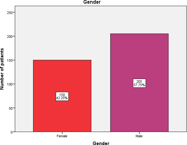
The collected data was then entered in Microsoft Excel spreadsheet and analysed using SPSS software (IBM SPSS Statistics, version 23). Frequency distribution and percentage were calculated for data summarization and presentation. The study sample consisted of 355 edentulous patients (150 females and 205 males) [Figure 1].
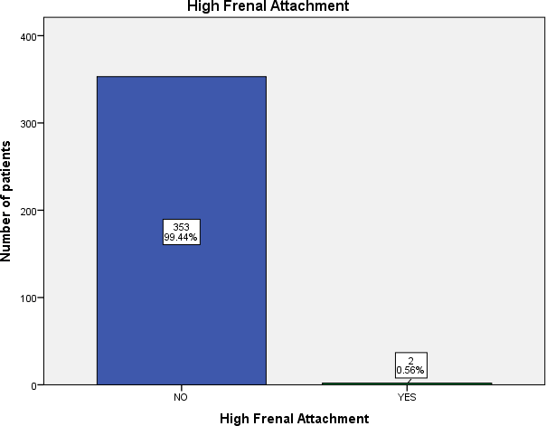
The mean age of the study population was 61.02 ± 9.85 years. Among the study subjects, 2 patients had high frenal attachment in the given population [Figure 2].
Figure 2: Bar chart showing distribution of high frenal attachment among study population. X-axis represents presence or absence of high frenal attachment and Y-axis represents the number of patients. Among 355 edentulous patients, 2 patients had high frenal attachment and 353 patients did not present with high frenal attachment.
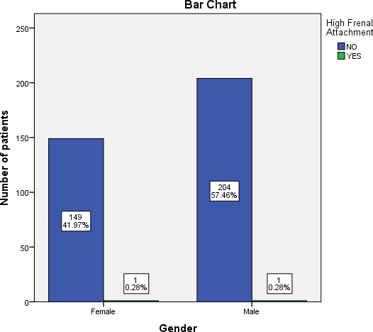
The association between gender and high frenal attachment using Chi-square test was done and found to be not statistically significant with p value of 0.824 [Figure 3].
Figure 3: Bar chart showing association between gender and high frenal attachment. X-axis represents gender and Y-axis represents the number of patients. Green denotes presence of high frenal attachment and blue denotes absence of high frenal attachment. Among 150 females, 1 patient had high frenal attachment and among 205 males, 1 patient had high frenal attachment. In both the gender, high frenal attachment was present in one patient. Association between gender and high frenal attachment was not statistically significant with the p value of .824 (Chi-square test)
both the gender, high frenal attachment was present in one patient. Association between gender and high frenal attachment was not statistically significant with the p value of .824 (Chi-square test)
The present retrospective study assessed the prevalence of high frenal attachment in edentulous patients.
Cilingir et al. [37] assessed the impact of frenulum height on strains in maxillary denture bases and suggested that the stress on the anterior midline of the maxillary complete denture increased with a higher labial frenulum. Al Jabbari [38] suggested that an adequate patient satisfaction with conventional complete dentures can be significantly increased after frenectomy. Also, Axinn recommended that where an unfavorable frenum is present, the technique of frenectomy plus free graft is perceived as an efficient, predictable procedure to improve the prognosis of a complete or partial denture. [39-42]
To the best of our knowledge, this is the first study to assess the incidence of high frenal attachment in edentutous patients.
Khursheed et al. assessed the prevalence of high frenal attachment in the Kurdish young population and found out that majority of the individuals had gingival and mucosal type frenal attachment. Also, Jindal investigated the variations in frenal morphology in the diverse population of Himachal Pradesh. The authors suggested that the normal frenum was most common. Out findings are in accordance with previous studies as the incidence of high frenal attachment is less. [43]
However, the findings cannot be generalized because of limited sample size and geographic limitation.
The number of patients who require complete dentures will continue to increase as a result of the increasing proportion of the adult population older than 55 years. Patient satisfaction with complete denture is related to the quality of life. [44] Hence, proper intraoral examination is needed to rule out unfavorable conditions which require preprosthetic surgery prior to the treatment. Our institution is passionate about high quality evidence based research and has excelled in various fields. [42–48] We hope this study adds to this rich legacy. [45-48]
Therefore, extensive research is needed in this field to find the variations in frenal morphology and attachment in edentulous patients.
Conclusion
Within the limitation of the present study, it was concluded that only 2 patients out of 355 edentulous patients presented with high frenal attachment. Therefore, the prevalence of high frenal attachment in edentulous patients was less in the given population.
REFERENCES
- Jyothi S, Robin PK, Ganapathy D, Others. Periodontal health status of three different groups wearing temporary partial denture. Res J Pharm Technol. 2017;10:4339–4342.
- Farmer JB. Preventive prosthodontics: maxillary denture fracture. J Prosthet Dent. 1983;50:172–5.
- Beyli MS, von Fraunhofer JA. An analysis of causes of fracture of acrylic resin dentures. J Prosthet Dent. 1981;46:238–41.
- Ranganathan H, Ganapathy DM, Jain AR. Cervical and incisal marginal discrepancy in ceramic laminate veneering materials: A SEM analysis. Contemp Clin Dent. 2017;8:272-8.
- Ariga P, Nallaswamy D, Jain AR, Ganapathy DM. Determination of correlation of width of maxillary anterior teeth using extra oral and intraoral factors in indian population: A Systematic Review. World Journal of Dentistry. 2018;9: 68-75.
- Duraisamy R, Krishnan CS, Ramasubramanian H, Sampathkumar J, Mariappan S, Sivaprakasam AN. Compatibility of non-original abutments with implants: Evaluation of microgap at the implant-abutment interface, with original and non-original abutments. Implant Dent. 2019;28:289-295.
- Selvan SR, Ganapathy D. Efficacy of fifth generation cephalosporins against methicillin-resistant Staphylococcus Aureus-A review. Res J Pharm Technol. 2016; 9:1815-1818.
- Ganapathy D, Sathyamoorthy A, Ranganathan H, Murthykumar K. Effect of resin bonded luting agents influencing marginal discrepancy in all ceramic complete veneer crowns. J Clin Diagn Res. 2016;10:ZC67–70.
- Subasree S, Murthykumar K, Dhanraj. Effect of aloe Vera in oral health-A review. Res J Pharm Technol. 2016;9:609.
- Ganapathy DM, Kannan A, Venugopalan S. Effect of coated surfaces influencing screw loosening in implants: A systematic review and meta-analysis. World Journal of Dentistry. 2017;8:496-502.
- Basha FYS, Ganapathy D, Venugopalan S. Oral hygiene status among pregnant women. Res J Pharm Technol. 2018;11:3099–102.
- Ajay R, Suma K, Ali SA, Kumar Sivakumar JS, Rakshagan V, Devaki V, et al. Effect of surface modifications on the retention of cement-retained implant crowns under fatigue loads: An in vitro Study. J Pharm Bioallied Sci. 2017;9:S154–60.
- Vijayalakshmi B, Ganapathy D. Medical management of cellulitis. Res J Pharm Technol. 2016;9:2067–70.
- Ashok V, Suvitha S. Awareness of all ceramic restoration in rural population. J Pharm Res. 2016;9:1691-1693.
- Ashok V, Nallaswamy D, Benazir Begum S, Nesappan T. Lip Bumper prosthesis for an acromegaly patient: A clinical report. J Indian Prosthodont Soc. 2014;14:279-282.
- Venugopalan S, Ariga P, Aggarwal P, Viswanath A. Magnetically retained silicone facial prosthesis. Niger J Clin Pract. 2014;17:260–264.
- Kannan A, Venugopalan S. A systematic review on the effect of use of impregnated retraction cords on gingiva. Res J Pharm Technol. 2018;11:2121–2126.
- Huang WJ, Creath CJ. The midline diastema: A review of its etiology and treatment. Pediatr Dent. 1995;17:171–179.
- Kahnberg KE. Frenum surgery. I. A comparison of three surgical methods. Int J Oral Surg. 1977;6:328–333.
- Coleton SH. Mucogingival surgical procedures employed in re-establishing the integrity of the gingival unit (III). The frenectomy and the free mucosal graft. Quintessence Int Dent Dig. 1977;8:53–61.
- Arvina R, Kumar B. Frenectomy with laterally displaced flap: A case report. Research Journal of Pharmacy and Technology. 2019;12:3669–3671.
- Ponnulakshmi R, Shyamaladevi B, Vijayalakshmi P, Selvaraj J. In silico and in vivo analysis to identify the antidiabetic activity of beta sitosterol in adipose tissue of high fat diet and sucrose induced type-2 diabetic experimental rats. Toxicol Mech Methods. 2019;29:276–290.
- Mathew MG, Samuel SR, Soni AJ, Roopa KB. Evaluation of adhesion of Streptococcus mutans, plaque accumulation on zirconia and stainless steel crowns, and surrounding gingival inflammation in primary molars: randomized controlled trial. Clin Oral Investig. 2020;24:3275–3280.
- Subramaniam N, Muthukrishnan A. Oral mucositis and microbial colonization in oral cancer patients undergoing radiotherapy and chemotherapy: A prospective analysis in a tertiary care dental hospital. J Investig Clin Dent. 2019;10:e12454.
- Girija ASS, Shankar EM, Larsson M. Could SARS-CoV-2-Induced Hyperinflammation Magnify the Severity of Coronavirus Disease (CoViD-19) Leading to Acute Respiratory Distress Syndrome? Front Immunol. 2020;11:1206.
- Dinesh S, Kumaran P, Mohanamurugan S, Vijay R, Singaravelu DL, Vinod A, et al. Influence of wood dust fillers on the mechanical, thermal, water absorption and biodegradation characteristics of jute fiber epoxy composites. J Polym Res. 2020;27.
- Thanikodi S, Singaravelu D Kumar, Devarajan C, Venkatraman V, Rathinavelu V. Teaching learning optimization and neural network for the effective prediction of heat transfer rates in tube heat exchangers. Therm Sci. 2020;24:575–581.
- Murugan MA, Jayaseelan V, Jayabalakrishnan D, Maridurai T, Kumar SS, Ramesh G, et al. Low velocity impact and mechanical behaviour of shot blasted SiC wire-mesh and silane-treated aloevera/hemp/flax-reinforced SiC whisker modified epoxy resin composites. Silicon Chem. 2020;12:1847–1856.
- Vadivel JK, Govindarajan M, Somasundaram E, Muthukrishnan A. Mast cell expression in oral lichen planus: A systematic review. J Investig Clin Dent. 2019;10:e12457.
- Chen F, Tang Y, Sun Y, Veeraraghavan VP, Mohan SK, Cui C. 6-shogaol, A active constiuents of ginger prevents UVB radiation mediated inflammation and oxidative stress through modulating NrF2 signaling in human epidermal keratinocytes (HaCaT cells). J Photochem Photobiol B. 2019;197:111518.
- Manickam A, Devarasan E, Manogaran G, Priyan MK, Varatharajan R, Hsu C-H, et al. Score level based latent fingerprint enhancement and matching using SIFT feature. Multimed Tools Appl. 2019;78:3065-85.
- Wu F, Zhu J, Li G, Wang J, Veeraraghavan VP, Krishna Mohan S, et al. Biologically synthesized green gold nanoparticles from induce growth-inhibitory effect on melanoma cells (B16). Artif Cells Nanomed Biotechnol. 2019;47:3297-3305.
- Ma Y, Karunakaran T, Veeraraghavan VP, Mohan SK, Li S. Sesame inhibits cell proliferation and induces apoptosis through inhibition of STAT-3 translocation in thyroid cancer cell lines (FTC-133). Biotechnol Bioprocess Eng. 2019;24:646-652.
- Ponnanikajamideen M, Rajeshkumar S, Vanaja M, Annadurai G. In vivo type 2 diabetes and wound-healing effects of antioxidant gold nanoparticles synthesized using the insulin plant Chamaecostus cuspidatus in albino rats. Can J Diabetes. 2019;43:82-89.
- Vairavel M, Devaraj E, Shanmugam R. An eco-friendly synthesis of Enterococcus sp.-mediated gold nanoparticle induces cytotoxicity in human colorectal cancer cells. Environ Sci Pollut Res Int. 2020;27:8166-8175.
- Paramasivam A, VijayashreePriyadharsini J, Raghunandhakumar S. N6-adenosine methylation (m6A): a promising new molecular target in hypertension and cardiovascular diseases. Hypertens Res. 2020;43:153-154.
- Cilingir A, Bilhan H, Baysal G, Sunbuloglu E, Bozdag E. The impact of frenulum height on strains in maxillary denture bases. J Adv Prosthodont. 2013;5:409-415.
- Al Jabbari YS. Frenectomy for improvement of a problematic conventional maxillary complete denture in an elderly patient: a case report. J Adv Prosthodont. 2011;3:236-239.
- Axinn S, Brasher WJ. Frenectomy plus free graft. J Prosthet Dent. 1983;50:16-19.
- https://www.researchgate.net/profile/Ranjdar_Talabani/publication/281627436_Prevalence_of_Labial_Frenum_Attachment_and_its_Relation_to_Diastemia_and_Black_Hole_in_Kurdish_Young_Population/links/55f08cbb08aef559dc46d44d.pdf.
- Jindal V, Kaur R, Goel A, Mahajan A, Mahajan N, Mahajan A. Variations in the frenal morphology in the diverse population: A clinical study. J Indian Soc Periodontol. 2016;20:320–323.
- VijayashreePriyadharsini J. In silico validation of the non-antibiotic drugs acetaminophen and ibuprofen as antibacterial agents against red complex pathogens. J Periodontol. 2019;90:1441-1448.
- Ezhilarasan D, Apoorva VS, Ashok Vardhan N. Syzygiumcumini extract induced reactive oxygen species-mediated apoptosis in human oral squamous carcinoma cells. J Oral Pathol Med. 2019;48:115-121.
- Ramesh A, Varghese S, Jayakumar ND, Malaiappan S. Comparative estimation of sulfiredoxin levels between chronic periodontitis and healthy patients - A case-control study. J Periodontol. 2018;89:1241–1248.
- https://link.springer.com/article/10.1007/s00784-020-03204-9.
- Sridharan G, Ramani P, Patankar S, Vijayaraghavan R. Evaluation of salivary metabolomics in oral leukoplakia and oral squamous cell carcinoma. J Oral Pathol Med. 2019;48:299-306.
- Pc J, Marimuthu T, Devadoss P. Prevalence and measurement of anterior loop of the mandibular canal using CBCT: A cross sectional study. Clin Implant Dent Relat Res. 2007.
- Ramadurai N, Gurunathan D, Samuel AV, Subramanian E, Rodrigues SJL. Effectiveness of 2% Articaine as an anesthetic agent in children: Randomized controlled trial. Clin Oral Investig. 2019;23:3543–3550.


 The Annals of Medical and Health Sciences Research is a monthly multidisciplinary medical journal.