Rare Anatomical Variant of the Inferior Vena Cava: Left Sided Inferior Vena Cava
2 Department of Surgery, Oncology National Institute, IbnSina University Hospital, Rabat, Morocco
Received: 13-Jul-2021 Accepted Date: Jul 20, 2021 ; Published: 30-Jul-2021
This open-access article is distributed under the terms of the Creative Commons Attribution Non-Commercial License (CC BY-NC) (http://creativecommons.org/licenses/by-nc/4.0/), which permits reuse, distribution and reproduction of the article, provided that the original work is properly cited and the reuse is restricted to noncommercial purposes. For commercial reuse, contact reprints@pulsus.com
Abstract
Anatomical variants of the inferior vena cava are important to know mainly for surgical treatment.They are mainly represented by the duplication of inferior vena cava, whereas left sided inferior vena cava is a rare variant.Imaging allows diagnosis of the variants, as they are usually discovered incidentally. We report the case of a 61 years old patient admitted for rectal cancer, to which a thoraco-abdominal extension examination CT-scan showed a left sided inferior vena cava.
Keywords
Anatomical; Variant; Inferior; Vena cava; Imaging
Introduction
The left sided inferior vena cava is a rare variant of the inferior vena cava due to an embryological anomaly of regression of the cardinal veins. It is mostly asymptomatic therefore usually diagnosed through imaging.
We report the case of a 61 years old patient admitted for rectal cancer, to which a thoraco-abdominal extension examination CT-scan showed a rare variant of the inferior vena cava: The left sided inferior vena cava.
Case Report
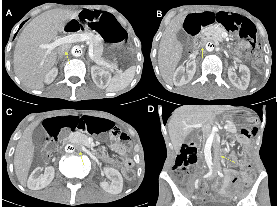
A 61 years old male patient, diagnosed with low rectal cancer, was admitted for a tumor extension examination through a thoraco-abdominal CT-scan. The abdominal enhanced CT-scan showed a left sided inferior vena cava [Figure 1].
The patient received concomitant chemoradiotherapy for his rectal cancer, followed by surgery (abdominoperineal amputation and pseudo-continent perineal colostomy), with a good follow up.
Discussion
The Inferior Vena Cava (IVC) is formed between the 6th and 8th gestational week, from 3 cardinal veins: Posterior cardinal, sub cardinal and supra cardinal veins. Their regression into one singular vein forms the inferior vena cava. [1]
Anomaly of regression of these veins results in anomalies of IVC such as duplication, interruption and transposition.
The duplication of IVC is the most commonanatomical variant one with an incidence of 0.2%-0.5% [1,2] whereas the left sided IVC represents a rare variant with a 0.1%-0.4% incidence according to a study published by Ang et al. between 2000 and 2011. [3] These variants are usually asymptomatic and discovered incidentally through imaging. [4,5] The left sided IVC is caused by the persistence of the left supra cardinal vein with regression of the right supra cardinal vein. [6] It is clinically asymptomatic, and can be of no clinical harm. Although sometimes the right renal vein can be compressed between the aorta and superior mesenteric artery causing a nutcracker syndrome. [6-10] Diagnosis is mostly done fortuitously through enhanced imaging either through a CTscan or an MRI. [4] These variants are of no serious impact but they are important to know and to be aware of especially for surgery in case of a tumor removal, lymph node dissection or transplant. [11]
Conclusion
Anatomical variants of the inferior vena cava, although asymptomatic, are important to know for surgical treatments.Imaging is the examination of choice for diagnosis.
REFERENCES
- Michael AG, Miguel C, Victor C, Gaetano C. Inverted nutcracker syndrome: A case of persistent hematuria and pain in the presence of a left-sided inferior vena cava. Sci World J. 2011;11:1031-5.
- Chen L, Jian Z, Quan Y, Yu Z, Hongguang Xu. A patient with left-sided inferior vena cava who received oblique lumbar interbody fusion surgery: A case report. J Med Case Rep. 2020;14(1):21.
- Wee Choen A, Terry D, Mark DS. Left-sided and duplicate inferior vena cava: A case series and review. Clin Anat. 2013;26(8):990-1001.
- Khalid E, Adil D, Mohamed D, Rachid A, Fethi M. La veine cave inférieure gauche et la grefferénale. Pan Afr Med J. 2019;34:109.
- Mototsugu M, Masaru F, Tomoyuki U, Keisuke M. Left-sided inferior vena cava. BMJ Case Rep. 2015;bcr:2015211268.
- Yasuhiro U, Hayato A, Takayuki O. Left-sided inferior vena cava with nutcracker syndrome. Clin Exp Nephrol. 2019;23(3):425-426.
- Nobuhisa T. Local advanced rectal cancer perforation in the midst of preoperative chemoradiotherapy: A case report and literature review. World J Clin Cases. 2017;23(7):326-339.
- Khalil El G. Rectal perforation after neoadjuvant chemoradiotherapy for low-lying rectal cancer. BMJ Case Rep. 2015;34:1-10.
- Bundgaard NS. Intraoperative tumor perforation is associated with decreased 5-year survival in colon cancer: A nationwide database study. Scand J Surg. 2017;26(8):890-901.
- Khan A. Rectal cancer perforation: A rare complication of neo-adjuvant radiotherapy for rectal cancer. Internet Journal of Oncology, 2010;36(1):1-10.
- Zbigniew B, Banaszkiewicz Z. Colorectal cancer with intestinal perforation-a retrospective analysis of treatment outcomes. Contemp Oncol (Pozn). 2014;89:1-25.


 The Annals of Medical and Health Sciences Research is a monthly multidisciplinary medical journal.