Recurrent Perianal Sinus in Young Girl Due To Pre‑sacral Epidermoid Cyst
- *Corresponding Author:
- Dr. Vinod Jain
B-41, Mahanagar Extension, Lucknow - 226 006, Uttar Pradesh, India.
E-mail: vinodjainkgmu@yahoo.co.in
Citation: Jain V, Misra S, Tiwari S, Rahul K, Jain H.Recurrent perianal sinus in young girl due to pre-sacral epidermoid cyst.Ann Med Health Sci Res 2013;3:458-60.
Abstract
Pre-sacral epidermoid cysts are rare development cysts resulting from dysembryogenesis mostly diagnosed in middle aged women. We report a case of pre-sacral epidermoid cyst presenting with recurrent perianal sinus in young girl. Generally pre-sacral epidermoid cysts are seen in adult age group but it is rare presentation in young age group. We report a rare case of presacral epidermoid cyst occurring in a young female.
Keywords
Coccygectomy, epidermoid cyst, Peri anal sinus, Young girl
Introduction
Pre-sacral epidermoid cysts are rare development cysts resulting from dysembryogenesis mostly diagnosed in middle aged women. Generally pre-sacral epidermoid cysts are seen in adult age group but it is rare presentation in young age group. Epidermoid cysts are benign unilocular lesions filled with clear fluid. These cysts are lined with stratified squamous epithelium.[1] There are three major complication of pre sacral developmental cysts. These are infections, bleeding and malignant degeneration3, which are the cause for intervention. Most of the time these are asymptomatic.[2] In our case it was the infective complication of epidermoid cyst which led to sinus formation.
Case Report
A young girl presented in our outpatient department with pus discharging from perianal sinus for the past 2 years. In addition, she was also having continuous dull pain in the lower abdomen along with constipation. Two years back, she was operated for this problem with misdiagnosis as fistula‑in‑ano. After 1 year period of remission, the sinus recurred. Then onwards, it persisted until the date of presentation.
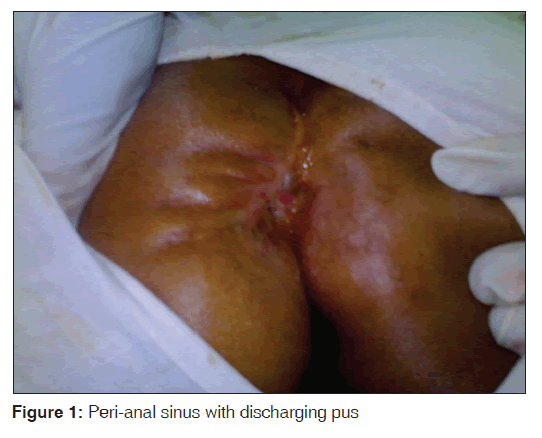
Local examination revealed a pus discharging from sinus at 6 o’clock position which was about 3 cm posterior to anal opening. Digital rectal examination revealed a bulge in the retrorectal area [Figure 1]. Compression over the swelling led to discharge of pus through the sinus opening.
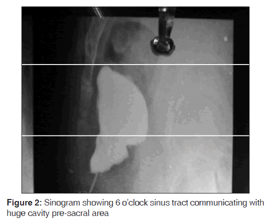
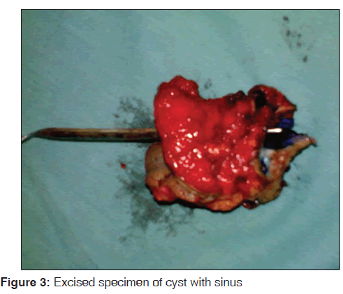
Laboratory tests were within normal limits. immunoglobulin G (IgG), immunoglobulin A (IgA), immunoglobulin M (IgM) antibodies along with polymerase chain reaction (PCR) for tuberculosis were negative. Ultrasonography of the abdomen was normal. Sinogram revealed a large pre‑sacral filling defect [Figure 2]. After pre‑operative assessment the cyst (8‑10 cm) was completely excised along with the sinus tract through posterior approach with coccygectomy [Figure 3]. Post‑operative period was uneventful.
Histopathology of excised specimen revealed it to be a case of epidermoid cyst lined with stratified squamous epithelium. No recurrence was noted after 2 years of follow‑up.
Discussion
Epidermoid cysts are benign unilocular lesions filled with clear fluid. These cysts are lined with stratified squamous epithelium.[1] Pre‑sacral epidermoid cysts are uncommon in young girls and presentation as perianal sinus is still rare. This lesion is mostly found in middle‑aged women and most of the time it is asymptomatic.[2]
There are three major complications of pre‑sacral developmental cysts, which are the cause for surgical intervention. These complications are infection, bleeding and malignant degeneration.[3] In our case, it was the infective complication of epidermoid cyst which led to sinus formation. Because of improper investigations and diagnosis, it was treated as fistula‑in‑ano in the past with no relief.
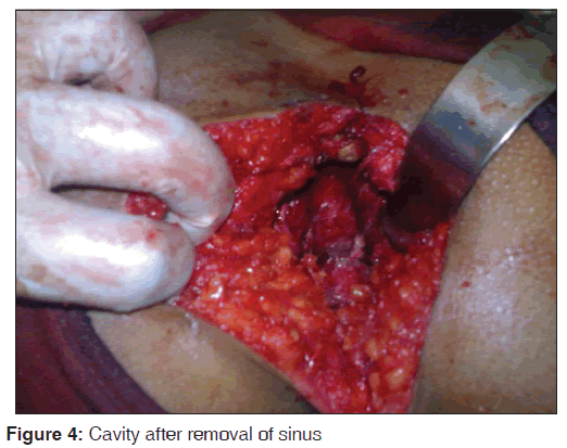
A case of incidentally found large pre‑sacral epidermoid cyst in young female was excised trans abdominally.[4] Other surgical options include a posterior approach, trans abdominal approach, combined approach or trans rectal approach.[5] We chose to operate through posterior approach in this case [Figure 4]. In a study, seven patients with retrorectal cysts have been misdiagnosed and treated as fistula‑in‑ano, pilonidal cyst, perianal abscess, lower‑back pain, post‑traumatic pain, post‑partum pain and proctalgia fugax before correct diagnosis.[6] Our case was also misdiagnosed and treated as fistula‑in‑ano elsewhere with no relief. Pre‑sacral epidermoid cysts have also been reported to be communicating with spinal CSF cavity,[7] but it is extremely rare.
Imaging with ultrasound is helpful in large pre-sacral epidermoid cyst with high clinical suspicion, but it is not always helpful to diagnose the disease, if the size of a cyst is small. In present case ultrasound imaging was more helpful as the size of cyst was large. We recommend ultrasound for all large pre-sacral epidermoid cyst.
Conclusion
Pre-sacral epidermoid cysts presenting as perianal sinus are uncommon in young girls. If misdiagnosed, it may be the cause of prolonged morbidity because of inadequate treatment. Therefore, high degree of clinical suspicion and proper investigations are needed in younger females presenting as perianal sinus.
Source of Support: Nil.
Conflict of Interest: None declared.
References
- Dahan H, Arriv� L, Wendum D, Docou le Pointe H, Djouhri H, Tubiana JM. Retrorectal developmental cysts in adults: Clinical and radiologic-histopathologic review, differential diagnosis, and treatment. Radiographics 2001;21:575-84.
- Leborgne J, Guiberteau B, Lehur PA, Le Goff M, Le N�el JC, Nomballais MF. Retro-rectal cystic tumors of developmental origin in adults. Apropos of 2 cases.Chirurgie 1989;115:565-71.
- Negro F, Mercuri M, Ricciardi V, Massari M, Destito C, Mafucci S, et al. Presacral epidermoid cyst: A case report. Ann Ital Chir 2006;77:75-7.
- Riojas CM, Hahn CD, Johnson EK. Presacral epidermoid cyst in a male: A case report and literature review. J Surg Educ 2010;67:227-32.
- Singer MA, Cintron JR, Martz JE, Schoetz DJ, Abcarian H. Retrorectal cyst: A rare tumor frequently misdiagnosed. J Am Coll Surg 2003;196:880-6.
- Nakamura S, Katagiri T, Majima A, Wakamatsu K, Tsubokawa T, Moriyasu N. Pre-sacral epidermoid cyst communicating with spinal CSF cavity (author?s transl). No Shinkei Geka 1979;7:389-95.
- Yang DM, Yoon MH, Kim HS, Oh YH, Ha SY, Oh JH, et al. Presacral epidermoid cyst: Imaging findings withhistopathologic correlation. Abdom Imaging 2001;26:79-82.


 The Annals of Medical and Health Sciences Research is a monthly multidisciplinary medical journal.