Sonographic Evaluation of the Splenic Length in Normal Pregnancy in a Tertiary Hospital in Southern Nigeria: A Pilot Study
- *Corresponding Author:
- Dr. Enighe W Ugboma
Department of Radiology, University of Port Harcourt Teaching Hospital, Port Harcourt, Rivers State, Nigeria.
E-mail: haugboma@gmail.com
Abstract
Background: The spleen is affected by the changes that occur in pregnancy. Ultrasound is the commonest imaging modality used in the evaluation of the abdominal organs in pregnancy; however, there is a paucity of information on the sonographic measurement of the splenic length in normal pregnancy in our environment. Aim: To establish sonographically the range of splenic length in normal pregnant women. Subjects and Methods: A prospective descriptive cross sectional study of the sonographic measurements of the splenic length was performed on 150 healthy normal pregnant women correlating this with the body mass index, gestational age and parity. Data were analyzed using software SPSS version 15 (SPSS Inc., Chicago, IL, USA). Correlations and variance between variables were calculated. P values 0.05 were considered as significant. Results: A mean splenic length of 10.0 cm (SD) 1.8 throughout pregnancy was obtained with a range of 9.7-10.3 cm. The splenic length significantly correlated positively with the body mass index (r = 0.006, P < 0.01) but not with parity (r = 0.94, P < 0.01), and gestational age (r = 0.31, P < 0.01). Conclusion: This study was able to establish a range of sonographic measurement of the splenic length for the locality.
Keywords
Normal pregnancy, Splenic length, South Nigeria, Ultrasound scan
Introduction
Normal pregnancy is a physiological state that alters the various systems of the body.[1] Enlargement of the spleen is seen in several pathological diseases which may occur in pregnancy requiring further work up.[2-4] Ultrasound scan, is the imaging modality of choice in pregnancy in that it is simple, reliable, noninvasive and repeatable, having advantage over other radiological imaging modalities such as computed axial tomography in that it uses sound energy not ionizing rays, thus is safe in pregnancy and can be used at any stage of pregnancy.[1] Its use is common in pregnancy in our setting. The sonographic measurement of the splenic length is important in the evaluation and follow up of patients with various pathologies,[5] as it is a good indicator and a quick method of evaluating the splenic size and used by majority of sonologists and sonographers in its evaluation.[6]
Review of literature shows that sonographic splenic size estimation has been done in various study population however there is a dread of information in the pregnant state in this environment.[7-11]
This paucity of published information on normative values for the splenic length in normal pregnancy in our environment, Nigeria and the West African sub region has necessitated this pilot study.
Subjects and Methods
A prospective descriptive cross sectional study on the sonographic evaluation of the splenic length in 150 normal pregnant women who were randomly chosen was carried out at the Radiology Department of University of Port Harcourt Teaching Hospital, Port Harcourt (UPTH), a 500 bed tertiary hospital in Rivers State, in the Niger Delta of Nigeria (with a catchment area of 23 local government areas and four states), q spanning through a six month period (February 2010 and July 2010). Ethical approval was obtained from the hospital’s Ethical committee. Informed consent was obtained from each participant. Those excluded from the study were those who had pre-existing suspected inflammations, metabolic, traumatic, collagen, or hemopoeitic diseases which could affect the splenic size.
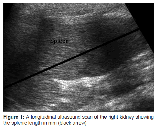
Real time gray scale ultrasound examination using Aloka 3500 machine fitted with a 3.5-5 MHz curvilinear transducer were used. Measurements were made in the supine position during deep inspiration. Splenic length (to the nearest millimetre) was obtained in the longitudinal section with the length taken from the dome of the spleen to the tip of the spleen through the splenic hilum [Figure 1]. All measured spleens were normal in position, shape and echotexture. Measurement was done by a single experienced researcher to reduce observer error. The intraobserver coefficient of variation for the measurement of splenic length was <10%.
Simple means and percentages were calculated, from which simple frequency tables, bar and pie chart were created. Data were analyzed using software SPSS version 15 (SPSS Inc., Chicago, IL, USA). Correlations and variance between variables were calculated. P values 0.05 were considered as significant.
Results
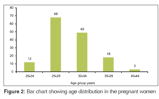
A total of 150 women took part in the study with their ages ranging from 20 to 41 years with average age of 29 years. The distribution is shown in Figure 2.
The parity ranged from 0 to 6 with women of parity 0 having the highest incidence 53/150 (35.3%) and those of parity 6, the lowest incidence 2/150 (1.3%).
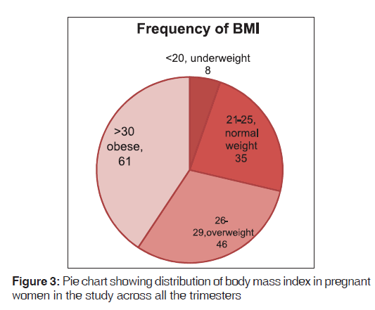
The body mass index (BMI) ranged from 19.5 to 54. with a mean of 29.45 [Figure 3].
Gestational age ranged from 9 to 40 weeks with an average of 28 weeks. Most of the women 93/150 (61%) were seen in the third trimester while only 9/150 (6%) were seen in the first trimester.
The mean splenic length throughout pregnancy was found to be 10.0 (1.8) cm with a range of 6.7-16.9 cm. The median value was 9.7 cm.
The lowest mean splenic length (7.98 cm (0.66)) was seen in the under weight group (BMI < 20). The highest values of splenic length were seen in those of BMI > 30 was 10.35 (2) cm. On the average there was a significant increase in the mean splenic length with increase in BMI with a significant positive linear correlation was seen between the BMI and splenic length [Table 1].
| Spleen | |
|---|---|
| BMI | |
| Pearson correlation | 0.223** |
| Sig. (2-tailed) | P<0.01 |
| N | 150 |
| GA | |
| Pearson correlation | 0.082 |
| Sig. (2-tailed) | 0.32 |
| N | 150 |
| Parity | |
| Pearson correlation | 0.006 |
| Sig. (2-tailed) | 0.94 |
| N | 150 |
| Spleen | |
| Pearson correlation | 1 |
| Sig. (2-tailed) | |
| N | 150 |
BMI: Body mass index, GA: Gestational age
Table 1: Correlations between the spleen, BMI, GA and parity
There was no significant steady increase in mean splenic length with increase in gestational age with the highest value, 10.08 (1.83) cm occurring in the third trimester and lowest 8.94 (0.89) cm in the first trimester.
There was also no significant correlation between the gestational age and splenic length [Table 1].
The highest value of the mean splenic length was 10.06 (1.91) cm in women of parity 2-4. On the average there was no significant increase in splenic length with increase in parity.
There was no significant correlation between parity and splenic length [Table 1].
Discussion
Ultrasound scan is the best imaging modality in the evaluation of the abdominal organs in pregnancy in that the examination is in real time, independent of organ function also, it is a noninvasive, nonionizing, easily available, cheap, safe, quick, and an accurate method for the measurement of the splenic length. The splenic length is quick to measure and gives an accurate estimation of the splenic size.[6]
There is a paucity of published information on the sonographic assessment of the splenic length in pregnancy although studies have been done in other groups such as in nonpregnant women, men, children, fetuses, and neonates.[2-4]
There was no significant increase in the splenic length throughout pregnancy (P < 0.01). The mean splenic length throughout pregnancy was seen to be 10.0 (1.8) cm. There is a paucity of established values for this environment and for normal pregnant women in general, however, compared to studies done elsewhere, this falls within the normal splenic length in the nonpregnant female.[5,7-9] Such studies include those by Spielmann, et al.,[10] were the average length of the spleen was found to be 10.3 ± 1.3 cm. Mittal[11] et al. obtained a slightly lower value of 9.34 ± 0.95 cm in females, while, Marco et al.[12] got a range of 8-11 cm with a median of 9.5 cm.
Weight gain is normal in pregnancy and with this, there is an increase in body mass index. In the absence of weight gain, poor pregnancy outcome is seen.[1] This weight gain is due to the weight of the fetus, placenta, membranes and liquor amni. In normal pregnancy, there is an increase in maternal weight which is due to the weight of the growing fetus, uterus, placenta, and membranes as well as retained fluids.[1] Swiet, et al.[13] postulated that weight gain of 9-12 kg is considered normal in pregnancy with weight gain occurring more during the first pregnancy. In course of the index pregnancy, weight gain accelerates in the third trimester with low weight gain associated with fetal growth restriction, low birth weight and a poor pregnancy out come.[10] The present study showed a significant positive linear correlation of the splenic length and body mass index. This finding is in concordance with Maymon et al.[14] where the splenic length was seen to correlate positively with the BMI. This was also seen in the nonpregnant female.[11] In pregnancy additional factors responsible for this could be explained by the fact that the increase in splenic length may be linked to other factors connected with the physiological state of pregnancy such as an increase in plasma flow.
There was no real significant difference in the splenic length across the various parities. Also there was no significant correlation of the splenic length with the parity. This could be due to the fact or confirm the fact that there is no significant increase in the splenic length as seen in this study during normal pregnancy thus the splenic length remains the same in subsequent pregnancies.
No significant correlations were seen between the splenic length and the gestational age. This was in contraindication to findings by Maymon et al.[14] who noticed a positive relation of the gestational age in a study done in Israel on the sonographic evaluation of the splenic length of 288 healthy pregnant women. This may be due to environmental and dietary factors. Also, this may be due to the small number of women recruited into the study across the various trimesters. More study is needed.
Conclusion
This study established a range of sonographic measurement of the splenic length in normal pregnancy in this environment, which may be used as a reference value for pregnant women with suspected spleenomegaly. The study also showed that the body mass index had a significant positive linear correlation with the splenic length but failed to establish a significant correlation between the parity and gestational age.
Limitation to this study, are the small population size, the inability to compare the pre and post pregnancy splenic length to the various factors as well as the pre and post weight gain/ BMI to the splenic length. Further investigation is needed to compare these factors as well as a larger study population is required, which might improve the precision of the estimates obtained.
Acknowledgments
We express our profound gratitude to the staff of the Radiology Department University of Port Harcourt Teaching Hospital. We, the authors have no potential conflicts of interest. The lead author has full access to all of the data in the study and takes responsibility for the integrity of the data and the accuracy of the data analysis
Source of Support
Nil.
Conflict of Interest
None declared.
References
- Beckmann C, Barzansky B, Herbert N, Ling F. Obstetrics and Gynaecology. 5th ed. Lippincott, Williams and Wilkins; Philadelphia, Pennsylvania 2005; p. 204.
- Morrafi D. Maternal changes during pregnancy. Geneva foundation for medical education. Available from: http:// www.gfmar.org. [Last cited on 2008].
- Kimberly D. Wallach EE. John Hopkins Manual of Gynaecology and Obstetrics. 2nd ed. Lippincott, Williams and Wilkins Philadelphia, Pennsylvania, 2002; p. 29.
- Tamayo SG, Rickman LS, Mathews WC, Fullerton SC, Bartok AE, Warner JT, et al. Examiner dependence on physical diagnostic tests for the detection of splenomegaly: A prospective study with multiple observers. J Gen Intern Med 1993;8:69-75.
- Lamb PM, Lund A, Kanagasabay RR, Martin A, Webb JA, Reznek RH. Spleen size: How well do linear ultrasound measurements correlate with three-dimensional CT volume assessments. Br J Radiol 2002;75:573-7.
- Niederau C, Sonnenberg A, Muller JE, Erckenbrecht JF, Scholten T, Fritsch WP. Sonographic measurements of the normal liver, spleen, pancreas and portal vein. Radiology 1983;149:537-40.
- Loftus WK, Chow LT, Metreweli C. Sonographic measurement of splenic length: Correlation with measurement at autopsy. J Clin Ultrasound 1999;27:71-4.
- Rodrigues Júnior AJ, Rodrigues CJ, Germano MA, Rasera Júnior I, Cerri GG. Sonographic assessment of normal spleen volume. Clin Anat 1995;8:252-5.
- Hosey RG, Mattacola CG, Kriss V, Armsey T, Quarles JD. Ultrasound assessment of spleen size in collegiate athletes. Br J Sports Med 2006;40:251-4.
- Spielmann AL, DeLong DM, Kliewer MA. Sonographic evaluation of spleen size in tall healthy athletes. AJR Am J Roentgenol 2005;184:45-9.
- Mittal R, Chowdhary DS. A pilot study of the normal measurements of the liver and spleen by ultrasonography in the Rajasthani population. J Clin Diagn Res 2010;4:2733-6.
- Marco P, Vincenzo M, Rosanna C, Ernesto S, Roberto M, Antonio S, et al. Measurement of spleen volume by ultrasound scanning in patients withthrombocytosis: A prospective study. Blood 002;99:4228-30.
- Swiet DM. Medical disorders in obstetric practice. 2nd ed. Hoboken, New Jersey Wiley-Blackwell; 1998. p. 203.
- Maymon R, Strauss S, Vaknin Z, Weinraub Z, Herman A, Gayer G. Normal sonographic values of maternal spleen size throughout pregnancy. Ultrasound Med Biol 2006;32:1827-31.


 The Annals of Medical and Health Sciences Research is a monthly multidisciplinary medical journal.