Subtrochanteric and Distal Femur Fractures in a Patient with Femoral Shaft Fracture Malunion and Knee Disarticulation: A Rare and Challenging Case Report
- *Corresponding Author:
- Dr. Pires RE
Department of the Locomotive Apparatus, Federal University of Minas Gerais, Avenida Professor Alfredo Balena, 190, Sala 193, Belo Horizonte, MG - 30130-100, Brazil.
E-mail: robinsonestevespires@gmail.com
This is an open access article distributed under the terms of the Creative Commons Attribution-NonCommercial-ShareAlike 3.0 License, which allows others to remix, tweak, and build upon the work non-commercially, as long as the author is credited and the new creations are licensed under the identical terms.
Abstract
This study aims to describe a rare and challenging case of a patient who presented ipsilateral subtrochanteric and distal femur fractures due to low‑energy trauma. The peculiarity of this case is the presence of femoral shaft fracture malunion and knee disarticulation in the same limb resulting from an accident suffered 30 years ago. The patient underwent femoral diaphyseal osteotomy and fixation of the subtrochanteric and distal femur fractures with a long cephalomedullary nail and distal femur locking plate, respectively. Despite the magnitude of the surgical procedure, all fractures healed, preserving the femoral length with the absence of infection and clinical complications. There was an improvement of the preinjury function attributed to the osteotomy of the femoral diaphyseal, which alleviated the anterior thigh discomfort.
Keywords
Amputation, Distal femur fracture, Femoral shaft fracture, Femur, Femur fractures, Fracture fixation, Fracture malunion, Fractures, Intramedullary nail, Knee disarticulation, Subtrochanteric fracture
Introduction
Femur fractures are present in the daily routine of the orthopedic surgeon. However, the association of proximal and distal fractures in the same femur greatly hinders the surgical management. The existence of a previous and severe injury, such as femoral diaphyseal fracture malunion with knee disarticulation in the same limb, makes the treatment truly demanding and with a high risk of complications.
This study reports the surgical management for this rare case and the treatment outcome after 1-year follow-up.
Ethical approval was granted by the local Ethics Committee, and the study was conducted according to the Declaration of Helsinki. Informed consent was obtained.
Case Report
Thirty years ago, a 20-year-old male patient suffered a car accident and presented closed femoral shaft fracture associated with a Gustilo-Anderson Type IIIC open fracture of the proximal tibia. He underwent primary knee disarticulation and conservative treatment of the femoral fracture.
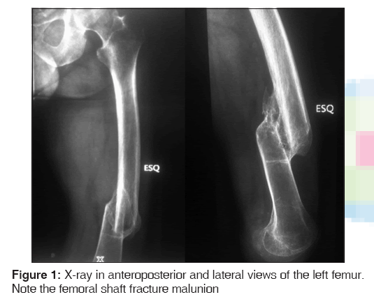
The patient developed femoral shaft fracture malunion that negatively affected rehabilitation with the below-knee prosthesis due to thigh discomfort [Figure 1].
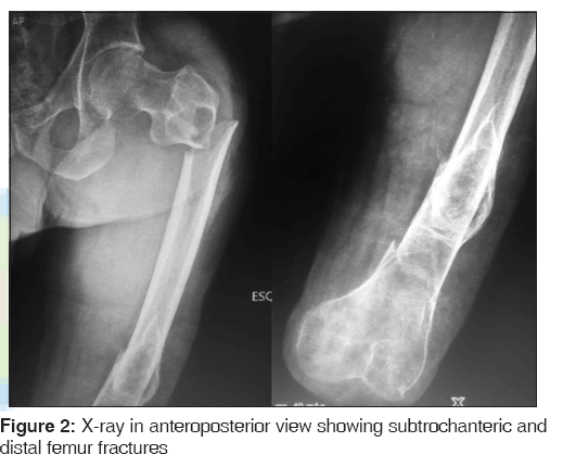
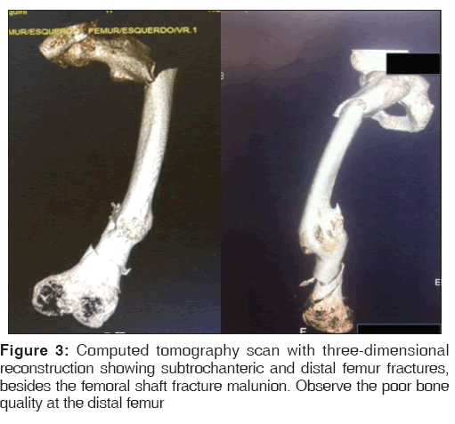
Thirty years later, the patient suffered a fall and sustained subtrochanteric and distal femur fractures in the same femur [Figures 2 and 3].
Surgical planning comprised femur shaft osteotomy preserving femur length, subtrochanteric fracture fixation with long cephalomedullary nail, and distal femur fixation with distal locking plate.
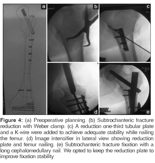
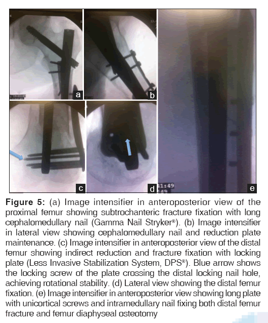
After failure of closed reduction attempt, we performed open reduction and provisional fixation of the subtrochanteric fracture using an anterior one-third tubular plate. Transverse osteotomy was performed at the diaphyseal malunion and a long cefalomedullary nail was used to fix the subtrochanteric fracture, diaphyseal osteotomy, and distal femur fracture. A locking plate was used to fix the distal femur fracture, enhancing the construction stability [Figures 4 and 5].
Figure 4: (a) Preoperative planning. (b) Subtrochanteric fracture reduction with Weber clamp. (c) A reduction one-third tubular plate and a K-wire were added to achieve adequate stability while nailing the femur. (d) Image intensifier in lateral view showing reduction plate and femur nailing. (e) Subtrochanteric fracture fixation with a long cephalomedullary nail. We opted to keep the reduction plate to improve fixation stability
Figure 5: (a) Image intensifier in anteroposterior view of the proximal femur showing subtrochanteric fracture fixation with long cephalomedullary nail (Gamma Nail Stryker®). (b) Image intensifier in lateral view showing cephalomedullary nail and reduction plate maintenance. (c) Image intensifier in anteroposterior view of the distal femur showing indirect reduction and fracture fixation with locking plate (Less Invasive Stabilization System, DPS®). Blue arrow shows the locking screw of the plate crossing the distal locking nail hole, achieving rotational stability. (d) Lateral view showing the distal femur fixation. (e) Image intensifier in anteroposterior view showing long plate with unicortical screws and intramedullary nail fixing both distal femur fracture and femur diaphyseal osteotomy
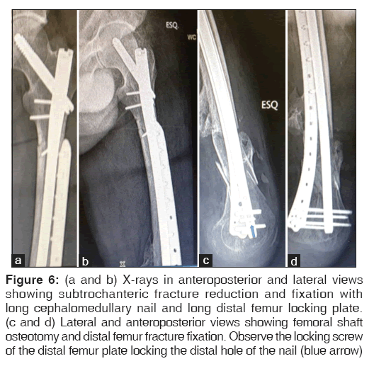
Immediate postoperative X-rays showing adequate fracture reduction and fixation [Figure 6].
Figure 6: (a and b) X-rays in anteroposterior and lateral views showing subtrochanteric fracture reduction and fixation with long cephalomedullary nail and long distal femur locking plate. (c and d) Lateral and anteroposterior views showing femoral shaft osteotomy and distal femur fracture fixation. Observe the locking screw of the distal femur plate locking the distal hole of the nail (blue arrow)
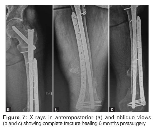
The patient evolved with no infection or clinical complications during the postoperative follow-up and started immediate physical therapy protocol. Partial weight bearing with the below-knee prosthesis was allowed after 3 months and total weight bearing after complete fracture healing (6 months postsurgery).
After 1-year follow-up, the patient presented no pain, absence of infection, complete fracture healing, and ability to walk without crutches [Figure 7].
Discussion
Femur fractures in amputated lower limb patients require high expertise to adequately manage surgical treatment. Difficulty achieving traction for indirect fracture reduction and poor bone quality are some of the major obstacles preventing satisfactory treatment outcomes.
Some authors have reported tricks to obtain fracture reduction in amputees using skeletal or skin traction applied to a fracture table to restore femoral length.[1-6]
In the present case, application of skin or skeletal traction to facilitate fracture reduction was impossible due to the presence of distal femur fracture. All maneuvers to obtain fracture reduction were performed using direct reduction with a Weber clamp proximally or by manual traction distally.
Another question concerning the association of proximal and distal femur fractures is whether to use one or two implants. When the proximal fracture is incomplete or minimally displaced, a reasonable decision is using a single implant to fix both fractures.[7] In the majority of cases, however, the rationale is fixing the fractures with two separate implants. In case of fixation failure, surgery revision is usually required in just one of the implants. In the present case, we decided to use two implants due to severity of fractures and high risk of complications.
Gao et al.[8] reported a case series of ipsilateral concomitant proximal extracapsular and distal femur fractures in no amputees. Functional outcomes were good in five cases, and fair and poor in one case each. According to the authors, proximal femoral nailing and distal femur locking plate could be a valuable and effective approach to handle ipsilateral concomitant proximal extracapsular and distal femur fractures.
Chen et al.[9] reported 15 cases of ipsilateral hip and distal femoral articular fractures caused by high-energy trauma in no amputees. Distal fracture patterns were more severe than proximal ones and the authors highlighted the high incidence of open joint injuries. An explanation for more complex fracture patterns at the distal femur could be the force injuring the distal femur directly and the proximal femur indirectly.
The uniqueness of this case is the presence of previous femoral shaft fracture malunion and knee disarticulation. The critical decision for this case was the choice of either only fixing the fractures or fixing the fractures plus associating femoral shaft osteotomy to correct femoral alignment, thereby alleviating anterior thigh discomfort and improving prosthesis adaptation. Regardless of the surgery magnitude, we decided to perform just a single surgical procedure which included fractures fixation and femoral osteotomy, consequentially preserving femur length.
A simpler treatment option for this case is fixation of the proximal femoral fracture with a short nail or a short plate, raising the amputation level until the femur malunion. Although the surgical procedure is minor, recent studies have shown that knee disarticulation leads to better functional outcomes than above-knee amputations due to more efficient lever arm for ambulation and preservation of muscle insertions.[10]
Another peculiarity of the present case was the use of a reduction plate to maintain the subtrochantercic fracture reduction. This is a helpful technical trick, especially for unstable fracture patterns. Nork et al.[11] first described the reduction plate technique to avoid valgus and extension of the proximal third of the tibia when nailing tibial fractures with the knee in flexion position. In this case, we decided to maintain the plate even after nailing the femur to improve fixation stability.
Conclusion
Femur fractures in amputees are challenging, even for experienced surgeons. The presence of femoral shaft fracture malunion and knee disarticulation caused by a previous trauma makes this case unique and defiant. We believe the surgical choice of performing femoral osteotomy and fractures fixation with independent implants was strongly decisive for the satisfactory treatment outcome with no complications after 1-year follow-up. Interesting surgical tricks were used in this case: reduction plate for the unstable subtrochanteric fracture and the locking screw of the plate distally locking the long nail to achieve rotational stability. Despite the singularity of this case, we believe the solution applied to the patient brings important messages that should be inserted in the treatment arsenal of the orthopedic surgeon.
Financial support and sponsorship
Nil.
Conflicts of interest
There are no conflicts of interest.
References
- Meena U, Meena R, Balaji S, Gaba S. Management of neglected femoral neck fracture in above knee amputated limb: A case report. Chin J Traumatol 2015;18:370-2.
- Anjum SN, McNicholas MJ. Innovative method of traction on fracture table in femoral neck fracture fixation in a below knee amputee. Inj Extra 2006;37:277-8.
- Mirdad T, Khan MR, Phil M, Kazarah Y. Fracture of the femur in amputation stumps. Ann Saudi Med 1997;17:638-40.
- Boussakri H, Alassaf I, Hamoudi S, Elibrahimi A, Ntarataz P, ELMrini A, et al. Hip arthroplasty in a patient with transfemoral amputation: A new tip. Case Rep Orthop 2015;2015:593747.
- Kandel L, Hernandez M, Safran O, Schwartz I, Liebergall M, Mattan Y. Bipolar hip hemiarthroplasty in a patient with an above knee amputation: A case report. J Orthop Surg Res 2009;4:30.
- Gamulin A, Farshad M. Amputated lower limb fixation to the fracture table. Orthopedics 2015;38:679-82.
- Ostrum RF, Tornetta P 3rd, Watson JT, Christiano A, Vafek E. Ipsilateral proximal femur and shaft fractures treated with hip screws and a reamed retrograde intramedullary nail. Clin Orthop Relat Res 2014;472:2751-8.
- Gao K, Gao W, Li F, Tao J, Huang J, Li H, et al. Treatment of ipsilateral concomitant fractures of proximal extra capsular and distal femur. Injury 2011;42:675-81.
- Chen CM, Chiu FY, Lo WH, Chuang TY. Ipsilateral hip and distal femoral fractures. Injury 2000;31:147-51.
- Penn-Barwell JG. Outcomes in lower limb amputation following trauma: A systematic review and meta-analysis. Injury 2011;42:1474-9.
- Nork SE, Barei DP, Schildhauer TA, Agel J, Holt SK, Schrick JL, et al. Intramedullary nailing of proximal quarter tibial fractures. J Orthop Trauma 2006;20:523-8.



 The Annals of Medical and Health Sciences Research is a monthly multidisciplinary medical journal.