The Fundamentals of Facial Skin Resurfacing for Practising Dental and Maxillofacial Surgeons: A Literature Review and Blueprint of Treatment Planning
2 Department of Oral and Maxillofacial Surgeon, Harvard Medical School,Harvard University, Boston, India
Received: 04-Nov-2022, Manuscript No. AMHSR-22-79315; Editor assigned: 07-Nov-2022, Pre QC No. AMHSR-22-79315 (PQ); Reviewed: 22-Nov-2022 QC No. AMHSR-22-79315; Revised: 03-Dec-2022, Manuscript No. AMHSR-22-79315 (R); Published: 10-Dec-2022
Citation: Tamhane A. The Fundamentals of Facial Skin Resurfacing for Practising Dental and Maxillofacial Surgeons: A Literature Review and Blueprint of Treatment Planning. Ann Med Health Sci Res. 2022;12:367-372
This open-access article is distributed under the terms of the Creative Commons Attribution Non-Commercial License (CC BY-NC) (http://creativecommons.org/licenses/by-nc/4.0/), which permits reuse, distribution and reproduction of the article, provided that the original work is properly cited and the reuse is restricted to noncommercial purposes. For commercial reuse, contact reprints@pulsus.com
Abstract
In the current era of corporate lifestyle and work culture, appearance has been a key factor in the scenario. Face, being that part of the body, only which can provide visual identity to an individual. Thus, the demand for facial skin health has been on a spike since the past decade. Environmental factors and certain pathological conditions have been deteriorating the surface quality and texture of the skin; in variably affecting the appearance of the individual. The medical science has shown evolution of various clinical procedures which aid in restoring the texture and quality of the facial skin. Skin resurfacing has been one of the key procedures in the skin rejuvenation. As a maxillofacial surgeon, we are predominantly equipped with deeper knowledge of the anatomy of the face and its structures. This provides us an edge over other physicians, in treating the histological, especially the dermal damages of the skin. The aesthetic and cosmetic facial procedures can be performed with minimal risk in an easily arrangeable set-up. It requires no general aneasthesia and can be taken up as walk-in treatment modality. Being non-surgical, there are no major systemic risks associated with the skin resurfacing procedures. The purpose of this particular article is to present various aspects of skin resurfacing with thorough detail, including the pre-procedure decisions such as patient selection and treatment selection to different procedures. The present literature review is an guiding aid for any maxillofacial surgeon who wants to step into the domain of facial aesthetics, and successfully treat the patient coming to the clinic with the cosmetic complaints.
Keywords
Corporate lifestyle; Appearance; Facial skin health; Texture; Skin resurfacing
Introduction
Appearance has been gaining popularity in the millennial era. We once believed in the fact that quotes ‘Never judge a book by its cover’; but in spite this being morally true has little practical significance. In the fast-moving world, there is a very little time for one to know a person inside-out, so ‘The first impression’ concept is valid significantly. ‘Face’ being the aspect that sets the first impression of an individual, has supreme value in overall appearance of a person.
Skin quality and texture gives a lot of attributes to the appearance of the face. Thus, it has become a prime concern to maintain or restore the health of the facial skin [2,3].
Hence considering the necessity of maintaining and restoring the skin health has been a concerned issue. Skin texture and quality can be glorified both conservatively and surgically. The surgical procedures being invasive, are associated with possible complications. Thus, patient masses prefer opting for non-invasive procedures, that aid to improve the skin health. Dermatologists and plastic surgeons have already ventured into providing access to the skin rejuvenating procedures, considering the increasing demand. Being a maxillofacial surgeon, invariably we are well versed with the anatomical and pathological aspects of the face. Thus, it becomes an area of prospect for the oral and maxillofacial surgeons to advance into.
Over the past few years, non-invasive skin rejuvenation procedures have shown hike in demand. A statistical data of 2017 in 2017, dermatologists in The United States Of America performed more than 9 million cosmetic treatments. This value was a 19% increase to that in 2016 [4]. According to another survey by ‘American Society Of Plastic Surgeons’, about total number of 15.9 Million non-invasive and minimally invasive skin rejuvenation procedures were performed only in the year 2018, that also was a 228% increase over year 2000 [5].
The rising demand for the skin rejuvenation has laid a new path for the oral and maxillofacial surgeons to explore. This creates an obvious need for the oral and maxillofacial surgeons to know, accumulate and understand the fundamentals of skin rejuvenation. ‘Skin rejuvenation’ in itself is a wide concept which involves different procedures. ‘Skin resurfacing’ is one of the technique to rejuvenate the aged or deteriorated skin. In the following review we provide a overview of the concept of skin resurfacing. The article includes description of anatomy of the skin and effects of aging, introduction to the concept of ‘Skin resurfacing’, diagnosis of the type of skin defect, treatment planning in the skin resurfacing procedures, concluding with different modalities of skin resurfacing and their categorisation. Hence with this particular article we intend to brief the maxillofacial surgeon’s community to the up-growing concept of ‘Skin resurfacing’[6,7].
Literature Review
Aging process and skin defects
In any contemplation over skin deterioration, it is important to demarcate between biological deterioration (chronological aging) and photoaging. The biologically aged skin clinically appears pretty distinct from the photoaged skin. The visible changes in biologically aged skin are paleness, laxity, deepening of expression lines, wrinkling, dryness and generalised thinning. Bruising is more common during biological aging and healing slows down. The photoaged skin is more yellowish in pigmentation with marked hyperpigmented areas, coarser and more rough in texture, more lax and more deeply wrinkled. As a thumb rule, individuals whose skin is biologically aged but photo protected appear younger than the individuals with photoaged skin. Table 1 gives us distinguishing aspects of biologically aged and photoaged skin [6,8].
| Table 1: Clinical appearance of biologically aged and photoaged skin. | ||
|---|---|---|
| Biologically aged skin | Photo aged skin | |
| Lax | Leathery | |
| Deepened expression lines | Dry | |
| Dry | Nodular and hypertrophied | |
| Overall thinning | Yellow | |
| _ | Telangiectasia | |
| _ | Deeper wrinkles | |
| _ | Accentuated skin furrows | |
| _ | Sags and bags | |
| _ | Variety of benign, premalignant and malignant neoplasms | |
Most pronounced changes in the biologically aged skin are seen in the epidermal layer, secondarily affecting the basal layer. The aging process is universal in all the areas of body than localised to face. General atrophy of the extra cellular matrix of the cells is depicted by a decrease in the number of the fibroblasts. There is reduction in the levels of collagen and elastin, impairment in the organization of the cells; all because of retardation in the process of protein synthesis. The histology presents with the flattening of the spiked interface of the dermis and epidermis. Also the skin barrier immunity is reduced, thermoregulation is affected, sensory perception is lowered due to the structural aging changes (Tables 2 and 3).
| Table 2: Regions of facial aging. |
|---|
| 1. Inherent changes within the skin |
| a) Epidermal changes |
| b) Skin tone and colour |
| c)Skin vasculature |
| d)Skin lesions |
| 2.Dynamic lines |
| a) Facial muscles acting on the skin |
| 3. Loss of tissue elasticity |
| a) Decreased elastin |
| b) Disarray of collagen bundles |
| 4. Effects of gravity |
| 5.Soft tissue and bone loss |
| a) Shifting of facial soft tissue |
| Table 3: Co-relation of histological change that is presented clinically with the particular component. | ||
|---|---|---|
| Visible change | Histological reason | |
| Thicker, irregular epidermis | Irregular stratum corneum | |
| Thinner dermis | a) Decreased elastin | |
| b) Thicker, irregular, disorganised collagen bundles | ||
| c) Decreased ground substance | ||
| Drier skin | Reduced appendages and glands | |
| Vessels become dilated and tortuous | Reduced microcirculation | |
| Lost laxicity | Thinner subcutaneous tissue | |
On the contrary to the biological aging, photoaging is characterised by hypertrophy. The sebaceous glands become enlarged, frequent neoplastic growths are encountered and the dermis of photoaged skin thickens with dilatation and derangement of blood vessels.The microvasculature also collapses and the vessels become more tortuous decreasing the blood supply to the skin and affecting its health. The number of hair follicles is also reduced and hair thinning is more prominent in the photoaged skin [6,9,10].
Acne is another skin pathology that causes surface defects and skin deterioration. As androgen levels in the body rise, the sebaceous glands become hypertrophic and increased sebum production leads to more severe form of acne. The excessive sebum trapped in hair follicle and bacterial flora sets the body’s immune response producing cystic lesions that involve the dermis, and when mechanically harmed produces scarring.
Skin pigmentation is another leading cause of skin quality deterioration. Melanocytes that produce melanin are responsible for the colour of the skin. Both hyper and hypo pigmentation are cause due to disruption in the count of melanocytes. The factors affecting the number of melanocytes are both extrinsic such as temperature, chemicals, and medications lead to atrophy of melanocytes and intrinsic such as hormones, genes affect melanocyte number. Skin resurfacing also caters the pigmentation issues of the skin [2,11].
Skin resurfacing and rejuvenation: Clarifying the terminologies
‘Skin rejuvenation’ is more over a generalised term which includes anything that is done to make skin look better. It includes non-medical amendments such as addition of more nutrient rich food and supplements to your diet that enhance the skin quality, application of topical products such as herbal substances or fruit pulps, applying vitamin serums etc. More technical procedures such as injectables, chemical peels, lasers are also included in the skin rejuvenation procedures [6,12].
Skin resurfacing is a specific set of procedures that are included under skin rejuvenation techniques, in which the built-up layers of debris and dead cells are cause to exfoliate leading to skins natural process of desquamation and collagen formation. All resurfacing treatments can address a wide variety of skin imperfections, including lines and wrinkles, discoloration, acne scars, and uneven skin tone. Skin resurfacing addresses either removal of debris or production of constituents of skin to smoothen the skin. The procedures can be mechanical (micro-dermabrasion), chemical (chemical peels) or radiation ( lasers). Thus while skin resurfacing is a more layman term, skin resurfacing is a structural amendment and a more professional approach. As an oral and maxillofacial surgeon proper knowledge of these resurfacing procedures, equips us with the skills of improving the appearance on an individual and have high aesthetic value [6,13,14].
The fundamentals of skin resurfacing
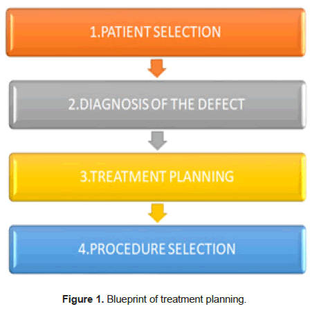
Skin resurfacing is a newer concept in the medical fraternity and thus there are various aspects other than the technicality of the procedures, that decide the outcome of the treatment and patient satisfaction. The entire process of skin resurfacing is a conceptually oriented aspect and thus the factors to be considered for the process are described sequentially in the flow chart below (Figure 1) [14,15].
Patient selection
While taking up any kind of aesthetic procedure, appropriate patient selection is a very important step. Patients undergoing cosmetic procedures most of the times approach the clinician with a consumer attitude. They expect immense results in inexpensive and manier times ‘doctor shop’. Because of variation in the individual expectations of the patients, demands and level of understanding it is important for the clinician to in detail educate the patient about realism of his expectations, varied treatment options, expected duration of the procedure, healing time and potential adverse events that may be associated with the procedure. The physician must also take in consideration the motivation level of the patient. Patients who approach for aesthetic treatments may be undergoing emotional dysfunction and lack of self-confidence. The patient may also be suffering from body dysmorphic disorder. Thus despite of the satisfactory treatment results, the patients with psychological and emotional imbalances will be not sufficiently satisfied post the treatment. Patient that should be ideally excluded from the selection are listed as follows:
• Pregnant women and lactating mothers
• Patients failing to keep follow up
• Patients demonstrating poor compliance with the topical skin conditioning regimen
• Patients with unrealistic expectations
• Medically compromised patients with co-morbidities
• Patients with psychological or emotional crisis
Diagnostic considerations
Patients diagnosis directly affects the time needed to complete the rejuvenation and resurfacing treatment. Scars and rhytides can be corrected relatively faster by modalities such as intralesional steroids, lasers, neurotoxins etc; while as pathologies such as melasma or other form of dermal melanosis takes much longer time and combination of modalities. The depth of treatment often guides the choice of required corrective procedure. Conditions limited to the epidermis respond adequately to the exfoliative procedures, where as conditions located within the dermis can be improved with the tightening or levelling procedures. Subcutaneous volume loss responds best to fillers, bio-stimulatory molecules or autogenous fat. Surgical removal of excess lax skin in also a rejuvenation modality for better results [6,14-16].
Treatment planning
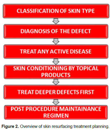
After identifying the patients skin type and diagnosis of the defect; formulating a comprehensive treatment plan is important for achiving both short term and long term goals. An overview of skin resurfacing treatment planning is established in the below flow chart (Figure 2).
Procedure selection
When choosing the procedure for a particular patient a clinician should have broader prospective considering skins overall quality, health and functional ability. There is no standardised selection criteria as such but various factors decide the procedure to be chosen. These factors include:
Skin type (Colour, thickness, oiliness, laxity and healing properties): Skin colour that is lighter is suitable for thermal procedures such as lasers, radiotherapy, ultrasound and chemical procedures. Darker skin tones give better results with non-thermal resurfacing procedures. Chemical procedures are more preferred for thicker skin types due to deeper penetration. Oily skins needed to be prophylactically treated with 5-month course of isotretinoin before an ablative or invasive procedure. For laxed skin chemical procedures penetrating to the depth of papillary dermis is more suitable. Furthermore, lost muscle laxity requires surgical approach for optimal cosmesis
According to nature of skin problem: Patients with underlying active disease such as acne, contact dermatitis, rosacea etc should not undergo a tightening or levelling procedure until the disease is adequately treated. Patients with epidermal and dermal melasma usually respond well to chemical peels as melanocytes are heat sensitive thus thermal procedures are avoided. Pathologies such as lentigines and ephelides are best treated with intense pulse light. Chronic photodamage is best treated by chemical peels as photodamaged skin is atrophic fragile and susceptible to scarring by ablative procedures. Dr. Zein Obagi skin stretch test is performed to identify the nature of skin problem.
Procedure selection according to mechanism of action: Resurfacing procedures vary in their mechanism of action. The procedures include: exfoliation, tightening, levelling, extracellular matrix stimulation and thickening, volume restoration. Table 4 enlists the depth of defect with required procedure.
| Table 4: Skin resurfacing procedure according to skin defect. | |
|---|---|
| Depth of penetration | Appropriate procedure to reach desired depth |
| Epidermis | Exfoliative or “false” peels |
| Papillary dermis (PD) | Tightening procedure, such as the ZO Controlled Depth Peel to the PD |
| Immediate reticular dermis (IRD) | Tightening procedure, such as the ZO Controlled Depth Peel to the IRD |
| PD, IRD, or upper reticular dermis (URD) | Combined tightening and levelling procedures, such as: |
| The ZO Designed Controlled Depth Peel when the URD is reached in small areas | |
| The ZO Controlled Depth Peel to the PD, IRD, and the URD, or | |
| The medium-depth ZO Designed Controlled Depth Peel when the URD is reached in large areas | |
| The combination of the ZO Controlled Depth Peel to the IRD followed by the CO2 fractional procedure to the URD | |
Non-surgical methods of skin resurfacing
Chemical Peels: Chemical peeling is one of the most preferred resurfacing techniques opted by both patient and the clinician. In comparison to newer modalities, chemical peels have high safety quotient and efficacy, easy to execute and cost effective. Chemical peels are actually varied acidic and basic compounds used to produce controlled and prophylactic skin injury which are classified as; superficial, medium depth and deep peeling agents according to their level of penetration in the skin. Superficial peels penetrate up to the epidermis, medium depth peels perforate to the papillary dermis and deep peels reach up to the mid reticular dermis [6,16,17].
Pre-treatment maximises the effect of the chemical peels and is indicated four to six weeks before the procedure. Tretinoin pre-treatment results in more uniform frosting and rapid reepithelisation. Hydroquinone is also indicated to decrease post treatment inflammatory hyperpigmentation. 10% glycolic acid administered topically also accelerates exfoliation. Immediately before the treatment the skin should be de-greased with acetone or alcohol. After this the chemical peeling agent can be applied to the face excluding the eyes, mouth and the alar facial groove as they possess risk of erosion.
Superficial chemical peels are considered for mild skin texture abnormalities and dyschromia. They have the advantage of minimal time of action and maximum effect. The effect of a superficial peel is confirmed by appearance of a white frost on the pink skin background.
Medium depth peels are indicated for treatment of the fine wrinkles. The action is confirmed on appearance of a white frost with erythematous strikethrough, indicating the changes in the papillary dermis. These peels require 3-7 days downtime during which patients may frequently experience erythema and swelling. [18,19]
Deep peels are aptly indicated for coarser wrinkles and deep acne scars. Due to deeper depth of penetration these peels should be administered under intravenous sedation or regional blocks. The action is confirmed with appearance of solid white frosting without any erythema.
The post-treatment should also be meticulously observed. After application of deep and medium peels patients are advised to moisturise frequently with petroleum-based jelly. Sun avoidance is also indicated up to two weeks after the procedure.
Laser resurfacing: Laser resurfacing induces an epidermal or dermal injury and helps treating the acne scars, fine wrinkles etc. The lasers can be of two types ablative and non-ablative (Table 4). Non ablative lasers such as Intense Pulse Light ( IPL), pulsed dye, Nd:YAG generate heat to induce injury and treating skin deformities without open wounds, while as ablative lasers create open wounds and induce bleeding. The thermal energy is hypothesised to stimulate the fibroblasts leading to collagen formation. The lasers can also be fractional or non-fractional. While fractional lasers injure the entire treated area, the nonfractional lasers injure the skin in columns with alternating unaffected areas [6,19].
Dermabrasion: Dermabrasion utilises an abrasive motorised wheel to create a mid-dermal wound. While as microdermabrasion usually employs an abrasive component usually a crystalline solid and a vacuum component. The central chamber of the delivery unit bursts the crystalline particles on the skin causing mechanical abrasion of dead cells. Newer epidermis is hence exposed. The abrasive particles and the dead cells are sucked out by means of the outer vacuum chamber [6].
Radiofrequency: Radiofrequency skin resurfacing works by transmitting the thermal energy to the reticular dermis, triggering the remodelling cascade of collagen and elastin formation and neovascularisation. It is a non-ablative technique that targets the dermis sparing the epidermis, thereby reducing the deep scars. It is indicated in correcting wrinkles, brow lifting nasolabial fold erection, jowls, marionette lines and skin laxity. Radiofrequency is contraindicated in the patients with healing disorders [6].
The modalities can be administered through monopolar, bipolar and unipolar devices. While monopolar frequency penetrates through all tissue layers, bipolar frequency is superficial in action. More common adverse reactions include swelling, numbness and bruising. The result outcomes can be seen in 6-12 months.
Ultrasound: Ultra sound uses a lower energy, micro focused energy to heat tissue through vibration and friction. This heating of the tissues produces small coagulation centres to induce remodelling of the reticular dermis. Unlike radiofrequency, ultrasound can treat deeper tissues without heating the dermal layers allowing transmission of higher energies to the superficial muscular aponeurotic system and platysma. During the treatment the tissues are under direct visualization and thus a pretty safe procedure. The procedure is performed by application of topical anaesthetic to relieve the caused pain. Focal depth is identified by visualizing on the monitor and the depth to be treated is calibrated for the linear array of ultrasound pulses to reach the target. The treatment time varies from 30 min-60 min per region [6].
Infra-red-light energy: Of the non-invasive skin resurfacing modalities, infra-red-light energy is the least studied. The procedure works by delivering infrared energy by a broadband light to create heat in the dermal layer and thus initiating remodelling of the collagen fibres. Unlike the other skin resurfacing modalities, infra-red energy doesn’t penetrate to the deeper subcutaneous tissues and hence is only indicated in superficial scarring and wrinkles [6,20,21].
Typically, this modality is not that painful and thus doesn’t require any anaesthetic aid. The procedure is carried out by thermally activating the treatment areas and then cooling by application of cold compresses. Due to the administration of heat, patient usually experience slight discomfort post treatment. Visible results are expected within 3-6 treatment cycles performed at 2–4-week interval. The results are variable to each skin type and the data is not well published on the complications encountered [22,23].
Microneedling: Micro needling is carried out by performing breaches in the epidermis by micron-sized needles causing points of injury in the form of micro punctures, which accelerate elastin and collagen production and deposition. The treatment is applicable to superficial epidermis only and can treat defects such as shallow rhytids. Micro needling induced drug delivery device is also available to directly let the pharmaceutical penetrate the target area [24,25].
Platlet rich plasma: Platelet Rich Plasma (PRP) is an autogenous plasma solution derived from patients own blood. Theory states that the PRP promotes wound healing by releasing secretory granules that contain growth factors to induce tissue generation and collagen production, increasing the skin thickness. PRP aids reverse aging and supplements for surgical grafts. PRP results in improved skin texture, elasticity, decreased rhytids, improved colour and reducing pigmentation. The ratio of PRP in the fat supplementation procedure varies from 2:1 to 10:1. The sites of PRP injections include nasolabial folds, malar and temporal region [26-28].
Most of the studies conducted for the PRP in facial resurfacing did not include control group, hence the results of these studies should be interpreted with caution. There are no standardised or evidence based protocols for using PRP in skin resurfacing that is why it is advised to the surgeon to have restricted and controlled use of PRP.
Conclusion
The With the growing awareness and demand for aesthetics and appearance conscious patients, skin resurfacing procedures are one of the avenues that a maxillofacial surgeon can take by educating himself about the available modalities and treatment planning. It is a less challenging and invasive field which attracts the patient class that belongs to higher economy zones. Thus, skin resurfacing practices have a high potential of business yield with lesser risks. Not many maxillofacial surgeons are versed with the modalities of skin rejuvenation and resurfacing and thus aesthetic facial surgical science is a newer domain to explore. The goal of writing this literature review was to provide a brief and comprehensive information bulk to the maxillofacial surgeons, that encourages them to more actively adapt to the facial aesthetic science and apply it in their clinical practice with well thought protocols, that bring out immense results without surgical intervention.
References
- Jones BC, Little AC, Burt DM, Perrett DI. When facial attractiveness is only skin deep. Perception. 2004;33:569-76.
[Crossref] [Google Scholar] [Indexed]
- Adamson PA, Galli SK. Rhinoplasty approaches: current state of the art. Arch Facial Plast Surg 2005;7:32-7.
- Little AC, Jones BC, DeBruine LM. Facial attractiveness: Evolutionary based research. Philos Trans R Soc Lond B Biol Sci. 2011;366:1638-59.
[Crossref] [Google Scholar] [Indexed]
- Houreld NN. The use of lasers and light sources in skin rejuvenation. Clin Dermatol. 2019;37:358-64.
[Crossref] [Google Scholar] [Indexed]
- Farber SE, Epps MT, Brown E, Krochonis J, McConville R, Codner MA. A review of nonsurgical facial rejuvenation. Plast Aesthet Res 2020;7:72.
- Obagi ZE. The art of skin health restoration and rejuvenation. The science of clinical practice, second edition. CRC Press. 2014.
- Lane MA, Mattison J, Ingram DK, Roth GS. Caloric restriction and aging in primates: Relevance to humans and possible CR mimetics. Microsc Res Tech. 2002;59:335–38.
[Crossref] [Google Scholar] [Indexed]
- Haisch A, Duda GN, Schroeder D, Gröger A, Geber C, Leder K, et al. The morphology and biomechanical characteristics of subcutaneously implanted tissue-engineered human septal cartilage. Eur Arch Otorhinolaryngol 2005;262: 993–97.
[Crossref] [Google Scholar] [Indexed]
- Choi YS, Park SN, Suh H. Adipose tissue engineering using mesenchymal stem cells attached to injectable PLGA spheres. Biomaterials 2005;26:5855–63.
[Crossref] [Google Scholar] [Indexed]
- Schmitz M, Graf C, Gut T, Sirena D, Peter I, Dummer R, et al. Melanoma cultures show different susceptibility towards E1A-, E1B–19 kDa- and fiber-modified replicationcompetent adenoviruses. Gene Ther. 2006;13:893–905.
[Crossref] [Google Scholar] [Indexed]
- Eppley BL, Kilgo M, Coleman JJ III. Cranial reconstruction with computer-generated hard-tissue replacement patient-matched implants: Indications, surgical technique, and long-term follow-up. Plast Reconstr Surg. 2002;109:864–71.
[Crossref] [Google Scholar] [Indexed]
- Steed DL, Group DU. Clinical evaluation of recombinant human platelet-derived growth factor for the treatment of lower extremity diabetic ulcers. J Vasc Surg. 1995;21:71–81.
[Crossref] [Google Scholar] [Indexed]
- Hom DB, Manivel JC. Promoting healing with recombinant human platelet-derived growth factor–BB in a previously irradiated problem wound. Laryngoscope 2003;113:1566–71.
[Crossref] [Google Scholar] [Indexed]
- Jakubowicz DM, Smith RV. Use of becaplermin in the closure of pharyngocutaneous fistulas. Head Neck. 2005;27:433–438.
[Crossref] [Google Scholar] [Indexed]
- Powell DM, Chang E, Farrior EH. Recovery from deep-plane rhytidectomy following unilateral wound treatment with autologous platelet gel: A pilot study. Arch Facial Plast Surg. 2001;3:245–250.
- Spielberger R, Stiff P, Bensinger W, Gentile T, Weisdorf D, Kewalramani T, et al. Palifermin for oral mucositis after intensive therapy for hematologic cancers. N Engl J Med. 2004; 351:2590–2598.
[Crossref] [Google Scholar] [Indexed]
- Chen W, Fu X, Ge S, et al. Ontogeny of expression of transforming growth factor-beta and its receptors and their possible relationship with scarless healing in human fetal skin. Wound Repair Regen 2005;13:68–75.
[Crossref] [Google Scholar] [Indexed]
- Becic F, Mulabegovi N, Mornjakovi Z, Kapić E, Prasović S, Becić E, et al. Topical treatment of standardised burns with herbal remedies in model rats. Bosn J Basic Med Sci. 2005;5:50–57.
[Crossref] [Google Scholar] [Indexed]
- Ferguson MW, O’Kane S. Scar-free healing: From embryonic mechanisms to adult therapeutic intervention. Philos Trans R Soc Lond B Biol Sci. 2004;359:839–50.
[Crossref] [Google Scholar] [Indexed]
- Matarasso A, Pfeifer TM, The Plastic Surgery Educational Foundation DATA Committee. Mesotherapy for body contouring. Plast Reconstr Surg. 2005;115:1420–1424.
[Crossref] [Google Scholar] [Indexed]
- American Society of Plastic Surgeons. Policy statement—mesotherapy. 2005.
- Devauchelle B, Badet L, Lengele B, Morelon E, Testelin S, Michallet M, et al. First human face allograft: Early report. Lancet. 2006;368:203–209.
[Crossref] [Google Scholar] [Indexed]
- Preminger BA, Fins JJ. Face transplantation: An extraordinary case with lessons for ordinary practice. Plast Reconstr Surg. 2006;118: 1073–74.
[Crossref] [Google Scholar] [Indexed]
- Alexiades-Armenakas M. Rhytides, laxity, and photoaging treated with a combination of radiofrequency, diode laser, and pulsed light and assessed with a comprehensive grading scale. J Drugs Dermatol 2006;5:731–738.
- Sadick NS, Trelles MA. Nonablative wrinkle treatment of the face and neck using a combined diode laser and radiofrequency technology. Dermatol Surg 2005;31:1695–1699.
[Crossref] [Google Scholar] [Indexed]
- Sewell C, Morris D, Blevins N, Barbagli F,Salisbury K. Quantifying risky behavior in surgical simulation. Stud Health Technol Inform 2005;111: 451–57.
- Challacombe BJ, Kavoussi LR, Dasgupta P. Trans-oceanic telerobotic surgery. BJU Int. 2003;92:678–80.
[Crossref] [Google Scholar] [Indexed]
- Takeuchi M, Tredget EE, Scott PG, Kilani RT, Ghahary A, et al. The antifibrogenic effects of liposome-encapsulated IFN-alpha2b cream on skin wounds. J Interferon Cytokine Res. 1999;19:1413–19.
[Crossref] [Google Scholar] [Indexed]


 The Annals of Medical and Health Sciences Research is a monthly multidisciplinary medical journal.