The use of Vitreous Cardio-Renal Biochemical Parameters as a Discriminant of Brackish Water Drowning
Received: 14-Jan-2024, Manuscript No. amhsr-24-131703; Editor assigned: 16-Jan-2024, Pre QC No. amhsr-24-131703 (PQ); Reviewed: 31-Jan-2024 QC No. amhsr-24-131703; Revised: 07-Feb-2024, Manuscript No. amhsr-24-131703 (R); Published: 14-Apr-2024
Citation: Agoro ES. The use of Vitreous Cardio-Renal Biochemical Parameters as a Discriminant of Brackish Water Drowning. Ann Med Health Sci Res. 2024;14:957-960.
This open-access article is distributed under the terms of the Creative Commons Attribution Non-Commercial License (CC BY-NC) (http://creativecommons.org/licenses/by-nc/4.0/), which permits reuse, distribution and reproduction of the article, provided that the original work is properly cited and the reuse is restricted to noncommercial purposes. For commercial reuse, contact reprints@pulsus.com
Abstract
Drowning is among the major causes of unintentional deaths with an annual estimate of about 372,000 as reported by the World Health Organization (WHO). Due to the empirical difficulties associated with discriminating true drowning from postmortem drowning, criminals now commit murder and disguise it as unintentional drowning. Drowning can occur in any kind of water body either artificial or natural. The type of water body associated with the drowning is crucial in the discrimination of postmortem drowning. Freshwater, brackish water, or saltwater drowning could present varied postmortem chemistries. Therefore, this study aimed to use some selected vitreous biomarkers and biochemical parameters in the discrimination of postmortem brackish water drowning. The sample size of this study was validated using Mead’s formula with a total of sixteen (16) rabbits employed. The study comprised four groups of four rabbits each. They include the Control Group (CD), the Chloroform Death group (CFD), the Postmortem Death Drowned group (PDD), and the Brackish Drowned Death (BDD). Statistical Package for Social Sciences (SPSS) program (SPSS Inc., Chicago, IL, USA; Version 18-21) was the choice statistical package. In a similar vein, One-Way ANOVA (LSD-Pos Hoc) was the choice tool for the vitreous data analysis. The study revealed elevated vitreous creatinine, uric acid, potassium, and chloride concentrations in death resulting from brackish water drowning. Other vitreous parameters were stable and could form part of the panel of laboratory investigations of brackish water drowning. The findings of this study could serve as a hallmark in defining brackish water drowning death.
Keywords
Brackish water drowning; Postmortem; Vitreous humor; Biochemical parameters; Biomarkers
Introduction
Drowning is a type of asphyxia instigated by the occlusion of the mouth and nose in a liquid either by submersion or immersion [1]. The estimated number of deaths resulting from drowning as of 2021 was 236,000 [2]. Drowning can occur in any kind of water body such as freshwater, brackish, or salt water. This study is centered on brackish water as the dearth of literature on this subject is quite conspicuous.
Brackish water is a type of natural water body with a salinity content higher than that of fresh water, and on the contrary lower than that of salt water [3]. Technically, brackish water contains between 0.5 and 30 grams of salt per liter. This is more often expressed as 0.5 to 30 parts per thousand (‰), with a specific gravity of between 1.0004 and 1.0226 [4].
Vitreous Humor (VH) is a clear interstitial gel-like fluid located between the lens and the retina of the eyeball [5,6]. It is found in both humans and animals with a similar physiological function of marinating the integrity of the eye. The VH makes up a fourthfifth of the volume of the eyeball and consists of inorganic ions such as Na+, K+, Cl2+, and Ca2+ [7,8]. The anatomical configuration makes some vitreous contents stable and less susceptible to autolytic changes [9,10]. This attribute is one of the major reasons VH is heavily patronized in forensic toxicology and postmortem chemistry.
Cardio-renal biomarkers are biochemical parameters that are either elevated, decreased, or stable in diseases or intoxication affecting both the heart and the kidney. These biomarkers are found majorly in the blood, but can also detected in other body fluids such as the vitreous [5,6]. The applications of these biomarkers are the same irrespective of the fluid or other matrices. Studies on the similarities of these biomarkers to diseases and intoxication investigations abound, though with different reference ranges [5,6].
In Nigeria, despite the paucity of data on drowning, the occurrences are rampant as reported in newspapers and eyewitness reports. This is of increasing concern in parts of the country predominated with water bodies especially in the Niger Delta region and Bayelsa State in particular. There are a handful of intentional drownings orchestrated and covered up as unintentional. This act is empirically difficult to resolve despite the utilization of autopsy procedures. In some instances, the autopsy procedure is not definitive and could create judicial gaps that create room for justice. This gap requires an empirical approach that is cheap, definitive, and complementary and could discriminate true drowning from that of postmortem. There are a handful of studies on the applications of vitreous humour chemistries in discriminating fresh-water drowning [1,6]. However, there is a paucity of research literature on that of brackish water. These twin gaps noted above are the fundamental basis of this study. This study was therefore aimed at using vitreous cardiorenal biochemical markers and electrolytes in discriminating death due truly to drowning from that postmortem drowning in brackish water.
Materials and Methods
Study area
This study was conducted in three different locations cutting across three Local Government Areas of Bayelsa State, Nigeria. The first location was domiciled at the Nembe River located in Nembe Town, where the water sampling was done. Animal breeding, artificial drowning, and sample collection were carried out at the animal house of the Federal University Otuoke. In a similar vein, the laboratory analysis was conducted at the Eniyimini Laboratories (eL) LTD located at Yenezue-gene Epie, Yenagoa, Bayelsa state [11].
Study population
A total of sixteen (16) rabbits were used for the study as validated by Mead’s resource equation [12]. The study comprised four groups of four rabbits each. They include the Control Group (CD), the Chloroform Death group (CFD), the Postmortem Death Drowned group (PDD), and the Brackish Drowned Death (BDD).
The control group rabbits were anesthetically euthanized mechanically without exposure to submersion. The CD died of chloroform inhalation intoxication. The PDD group was sacrificed before submersion, whereas the BDD group died of brackish water submersion. The study duration was pegged at twelve (12) hours to mimic the optimal time for the recovery of drowned cases.
Ethical approval
The ethical approval was granted by the Directorate of Research and Quality Assurance of the Federal University Otuoke, Bayelsa State. The laid down policy on the care of animals for research was stringently adhered to [13].
Research design/selection criteria
The research design used for the study was drawn from that of Adias et al., [14]. Only rabbits deemed to be fit by the University Veterinarian were recruited into the study. The sex of the animal used for the study was strictly male albino rabbits.
Vitreous humor collection
The vitreous humor samples were collected by the method of Coe [15]. Briefly, using a 5 ml syringe and a needle, a scleral puncture was made on the lateral canthus, and the total extractable vitreous humor was aspirated from the eye. Adequate care was taken to gently aspirate the fluid to avoid tearing any loose tissue fragments surrounding the vitreous chamber. On average 1.0 mL was collected from each rabbit’s eye. Only crystal-clear liquid free of tissue contaminants and fragments was used in the study.
The samples were collected within 30 minutes after the termination of the study into fluoride oxalate tubes for determination of glucose concentration and plain containers for other cardio-renal biochemical parameter analysis.
Laboratory procedure
Ion Selective Electrode (ISE) (analyzer ISE 4000-Germany) was used for the estimation of the vitreous electrolytes [16]. Sodium, potassium, chloride, bicarbonate, and calcium were the chosen vitreous electrolytes used for the study. Vitreous Creatine Kinase (CK-MB) and Lactate Dehydrogenase (LDH-P) activities were assayed quantitatively using the AGAPPE Diagnostics kit (Switzerland). Vitreous urea and creatinine were estimated using the diacetyl monoxime and Jaffes methods respectively [17]. In a similar vein, vitreous uric acid concentration was estimated quantitatively by the uricase method using the AGAPPE Diagnostics kit (Switzerland).
Analysis of date
The data generated from the study were analyzed with the aid of the Statistical Package for Social Sciences (SPSS) program (SPSS Inc., Chicago, IL, USA; Version 18-21) and Microsoft Excel. One-way ANOVA (Post Hoc) was used in comparing the means of the various biochemical parameters of the various groups of the study. Tables and charts were used for the presentation of various findings.
Results and Discussion
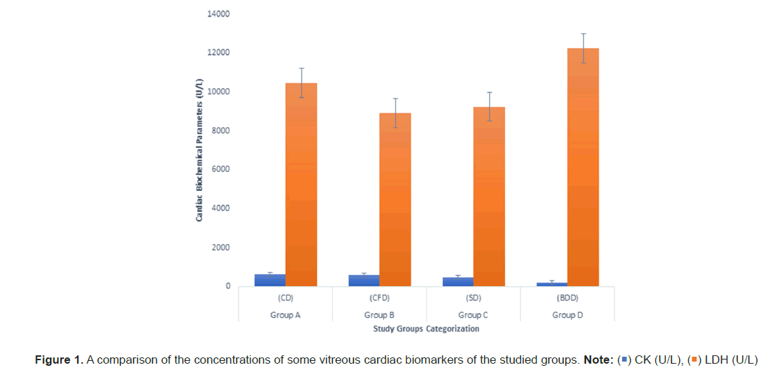
Table 1 shows a significant increase (P<0.05) in vitreous potassium concentration in the PD and BDD when compared to other groups. In a similar vein, vitreous chloride significantly increased in the BDD group when compared to the other groups. The concentrations of calcium and bicarbonate decreased significantly in the CFD group when compared to the CD. Table 2 revealed a significant difference in vitreous uric acid concentration in the BDD when compared to other groups. However, vitreous creatinine increased significantly in the BDD group when compared to the PD. Figure 1 on cardiac biomarkers exhibited no significant difference across all the groups.
| Electrolytes | Group A (CD) | Group B (CFD) | Group C (PD) | Group D (BDD) | F-test | P value |
|---|---|---|---|---|---|---|
| Sodium (mmol/L) | 152.863 ± 34.950 | 155.141 ± 43.815 | 200.346 ± 32.985 | 174.485 ± 30.691 | 1.124 | 0.395 |
| Potassium (mmol/L) | 12.479 ± 1.728 | 13.158 ± 0.912. | 23.581 ± 1.085a,b | 20.369 ± 0.861a,b | 62.246 | 0 |
| Chloride (mmol/L) | 74.0479 ± 5.518 | 85.355 ± 50546 | 76.061 ± 13.188 | 91.874 ± 2.891a,b | 3.192 | 0.085 |
| Bicarbonate (mmol/L) | 31.677 ± 4.582 | 19.572 ± 5.930a | 28.779 ± 6.924 | 39.764 ± 13.257 | 2.978 | 0.096 |
| Calcium (mmol/L) | 0.904 ± 0.356 | 0.364 ± 0.178a | 1.152 ± 0.248 | 0.792 ± 0.507 | 2.725 | 0.114 |
Table 1: A comparison of the mean concentrations of some vitreous electrolytes in the various groups.
| Parameters | Group A (CD) | Group B (CFD) | Group C (PD) | Group D (BDD) | F-value | P-value |
|---|---|---|---|---|---|---|
| Urea (mmol/l) | 10.719 ± 3.462 | 17.095 ± 3.656 | 23.572 ± 18.574 | 11.542 ± 5.348 | 1.063 | 0.417 |
| Uric acid (mmol/l) | 0.059± 0.008 | 0.241 ± 0.322 | 0.044 ± 0.029 | 8.246 ± 2.884 a,b,c | 23.226 | 0 |
| Creatinine (mmol/l) | 219.29 ± 78.112 | 383.576 ± 120.011 | 40.373 ± 9.908 | 709.239 ± 289.099c | 9.313 | 0.005 |
Table 2: Correlation of leg varicose veins and other study variables Age factor, work history, education, body mass index.
The study revealed a significant rise in vitreous potassium concentration in the PDD and BDD when compared to other groups (Table 1). In a similar vein, vitreous chloride concentration significantly increased in the BDD group when compared to the other groups. On the contrary, there was a fall in the concentration of vitreous calcium and bicarbonate concentrations in the CFD when compared to the CD.
The increase in the concentrations of vitreous potassium in the PDD and BDD could be attributed to the brackish water effect as against the Postmortem Interval (PMI) effect or cardiac arrest as portrayed in literature. Postmortem interval (PMI) is known to alter vitreous potassium concentrations as the interval increases [15,18]. The PMI of CD and CFD were longer than that of PDD and BDD but presented lower vitreous potassium concentrations. This affirmed the brackish water effect rather than the PMI effect.
The increased concentration of vitreous chloride observed in the BDD as against other groups also points to the brackish water effect. The increase could be attributed to hyperchloraemia resulting from brackish water intoxication. Chloride concentration is quite high in brackish water and could have infiltrated the vitreous during the drowning process. This study agrees with the findings of Allan [19], and that of Mahmoud et al., [20]. The decrease in calcium and bicarbonate concentrations in the CFD group compared to other groups could be attributed to the mechanism of action of chloroform. This observation is similar to a case study carried out by Cătălina [21] on chloroform inhalation.
Furthermore, this study revealed a significant increase (P<0.05) in vitreous creatinine and uric acid concentrations in the BDD when compared to other groups (Table 2). The empirical fact of these discriminatory alterations in the BDD could be due to muscular contractions resulting from the extreme aggregations during the drowning process, rather than renal insufficiency or compromise. Creatinine, urea, and uric acids are markers of renal insufficiency [22]. In this case, creatinine concentration singly increased as against the synergistic increase with urea. This affirms the muscular contraction stance of the authors.
Vitreous cardiac markers were not affected by brackish water drowning, or any other type of mortality as posited in this study (Figure 1). The cardiac markers commonly used in Nigeria for the assessment of cardiac fidelity are CK-MB and LDH. The stability of these parameters give credence to the exclusion of cardiac collapse as the primary cause of death resulting from brackish water drowning. The study has implicated some vitreous biochemistry parameters that could corroborate death resulting from brackish water drowning with other methods.
Conclusion
The study revealed elevated vitreous creatinine, uric acid, potassium, and chloride concentrations in death resulting from brackish water drowning. Other vitreous parameters were stable and could form part of the panel of laboratory investigations for brackish water drowning. The finding of this study could serve as a hallmark in defining brackish water drowning death. It has also opened up a robust discourse for scientific interactions on the importance of vitreous chemistry in confirming brackish water drowning and other kinds of drowning.
References
- Agoro ES, Ikimi CG, Edidiong T. The use of vitreous renal chemistries in the discrimination of postmortem freshwater drowning. Toxicology Research and Application. 2021;5:1–6.
- World Health Organization (WHO). Preventing drowning: An implementation guide online. 2021.
- Klaassen K, Borman H, Klenke T, Liebezeit G. The impact of hydrodynamics and texture on the inflitration of rain and marine waters into sand bank island sediments. Senckenbergiana maritima, 2008;38:163–171.
- Yvana D, Ahdab DR, John HL. Brackish water desalination for greenhouses: Improving groundwater quality for irrigation using monovalent selective electrodialysis reversal. Journal of Membrane Science. 2020; 610:118072.
- Agoro ES. Vitreous humor biochemical indicators of acute co intoxication associated deaths: A review of the literature. Arab Journal of Forensic Sciences and Forensic medicine. 2020a;2:58-61.
- Agoro ES, Ben-Wakama EN, Alabrah PW. Review on chronic impacts of carbon monoxide intoxication on some routine vitreous and blood investigations. Toxicol Forensic Med Open J. 2020b; 5:16-25.
- Susan S, Neil RB and Henry G. Gray’s anatomy: The anatomical basis of clinical practice. 40th edn. London: Churchill Livingstone. 2008.
- Jaffe FA. Chemical postmortem changes in the intraocular fluid. J Forensic Sci. 1962; 231-23. [Crossref]
[Google Scholar] [PubMed]
- Negrusz A, Cooper G. Postmortem toxicology. Clarke’s analytical forensic toxicology. 2nd edn. London: PhP Pharmaceutical Press. 2013:189–213.
- Agoro ES, Azuonwu O, Abbey SD. The forensic application of microbiological quality of the vitreous humor on the investigation of acute and chronic carbon monoxide poisoning. GSL J Forensic Res. 2017a; 1:1-4.
- Agoro ES, Chinyere GC, Akubugwo EI. Emerging concept of vitreous concentrations of proteins and lipids as discriminant of fresh water drowning death. Journal of Forensic and Legal Medicine. 2020c;73.
[Crossref] [Google Scholar] [PubMed]
- Kirkwood J, Robert H. The ufaw handbook on the care and management of laboratory and other research animals. Wiley-Blackwell. 2010;29.
- Benjamin A, Jean L. Legislative history of the animal welfare act. 2016.
- Adias TC, Ajugwo AO, Erhabor T, Nyenke, CU. Effect of pumpkin extract (telfairia occidentalis) on routine haematological parameters in acetone-induced oxidative stress albino rats." American Journal of Food Science and Technology. 2013: 67-69.
- Coe JI. Vitreous potassium as a measure of the postmortem interval: An historical review and critical evaluation. Forensic Sci Int. 1989; 42:201-13.
- Bolarin DM, Azinge EC. Chemical pathology laboratory tests in pregnancy. Journal of Medical Laboratory Science. 2010;19:3-13.
- Michael LB, Edward PF, Larry ES. Clinical chemistry: Principles, procedures, correlations. Philadelphia: Lippincott Williams & Wilkins.1992;100-200.
- Agoro ES, Okoye FBC, Onyenekwe CC, Azuonwu, O, Ebiere NE. Extrapolation of three hourly post-mortem intervals using some vitreous chemistry parameters. Journal of Forensic Research, 2017b;8:1-5.
- Allan C. Post-mortem vitreous sodium and chloride levels–a biochemical test for salt water drowning? Pathology. 2011;43:72.
- Mahmoud ME, Hadeer HR, Mohamed FH, Fathy RS. Biochemical markers and pathological features of postmortem time interval distinguishing freshwater and saltwater drowing induced death in albino rats. Medical Journal of Scientific & Technical Research. 2019;20:14713-14720.
- Cătălina L. Lethal complications after poisoning with chlorofor m-case report and literature review. Human and Experimental Toxicology. 2010; 29:615–622
[Crossref] [Google Scholar] [PubMed]
- Jeremiah ZA, Uko EK, Buseri FI, Adias TC. Baseline iron status of apparently healthy children in port harcourt, nigeria. Eur J Gen Med. 2007;4:161-4.


 ) CK (U/L), (
 ) LDH (U/L)


 The Annals of Medical and Health Sciences Research is a monthly multidisciplinary medical journal.