Total Maxillary Arch Distalization with Bone Screws in Infrazygomatic Crest: A Clinical Cephalometric Study
Received: 07-Jul-2023, Manuscript No. AMHSR-23-105310; Editor assigned: 10-Jul-2023, Pre QC No. AMHSR-23-105310 (PQ); Reviewed: 24-Jul-2023 QC No. AMHSR-23-105310; Revised: 07-Dec-2023, Manuscript No. AMHSR-23-105310 (R); Published: 15-Dec-2023
Citation: Patil PS, et al. Total Maxillary Arch Distalization with Bone Screws in Infrazygomatic Crest: A Clinical Cephalometric Study. Ann Med Health Sci Res. 2023;13:896-902
This open-access article is distributed under the terms of the Creative Commons Attribution Non-Commercial License (CC BY-NC) (http://creativecommons.org/licenses/by-nc/4.0/), which permits reuse, distribution and reproduction of the article, provided that the original work is properly cited and the reuse is restricted to noncommercial purposes. For commercial reuse, contact reprints@pulsus.com
Abstract
Objective: To evaluate the skeletal, dental, and soft tissue cephalometric changes produced by total arch distalization of maxilla using infrazygomatic crest bone screw as anchorage.
Methods: A prospective sample of 13 patients (5 females and 8 males) who underwent total maxillary arch distalization treatment with infrazygomatic crest bone screw as anchorage system. Lateral cephalograms were obtained at the beginning of Treatment (T0) and after class II molar correction (T1). To compare cephalometric changes, paired t-test was used. A significance level of 5% was used.
Results: Class I molar and class I canine relation was achieved in all patients. The IZC bone screw provided molar distalization of 4.17 mm and intrusion of 1.25 mm with distal tipping of 3.7°. Significant Maxillary incisor retraction of 4.58 mm and retroclination of 7.91° was observed. The upper lip in relation to the E line was retracted by 1.1 mm.
Conclusion: Effective skeletal, dental, and soft tissue treatment results were achieved using IZC bone screw for total maxillary arch distalization.
Keywords
Total maxillary arch distalization; Infrazygomatic crest; Bone screw; Temporary Anchorage Devices (TADs); Skeletal maxillary
Introduction
Class II malocclusion is one of the most prevalent types of malocclusion in orthodontic practice, being observed in 38% to 50% of patients [1]. It has been related to less favourable perceptions of facial and dental esthetics, contributing negatively to the quality of life and self-esteem of patients. The treatment for patients with class II division 1 malocclusion with skeletal maxillary excess can be either surgical or camouflage. Surgical correction is considered only in severe form of class II malocclusion. On the other hand, in camouflage treatment, the upper premolars are extracted to resolve crowding, retract the incisor, and provide class I canine relation [2]. Camouflage treatment can also be done using is molar distalization. However, each option requires considerable cooperation of patients to fulfill their real goals [3].
To overcome dependence on patient compliance, Temporary Anchorage Devices (TADs) were introduced placed between the roots of posterior teeth to promote the retraction of anterior teeth [4,5]. However, mini-screw have some major problems such as a high rate of failure, interference with the path of tooth movement, and impingement on the roots of adjacent teeth [6], Recently introduced techniques such as maxillary arch distalization by bone screws have overcome unwanted side effects are more effective and give good results. The Infrazygomatic Crest (IZC) has been an obvious choice for bone screw insertion because of the thickness of the cortical plate and its distance from the dental arch. Its ability to be placed high up in the maxillary sulcus, i.e., in the Infrazygomatic (IZ) area, allows unobstructed tooth movement during retraction [7].
There were no prospective studies evaluating the effects of maxillary dentition distalization with bone screw implanted in the IZC. Therefore, the objective of this study was to evaluate the efficacy of IZC bone screws and skeletal, dental and soft tissue cephalometric changes produced by total arch distalization of maxilla using infrazygomatic crest bone screw as anchorage.
Materials and Methods
The present study was a prospective clinical cephalometric evaluation study. It involved 13 patients reporting to the department of orthodontics and dentofacial orthopedics for orthodontic treatment at Bharati Vidyapeeth dental college and hospital, Sangli, Maharashtra. Case selection for the study was done based on the following criteria:
• Patients with the age of 12 and above.
• Patients with angle’s class II division I malocclusion.
• Protrusion of maxillary anterior teeth (3 mm ≤ over jet ≤ 9 mm.
• Patient having all permanent teeth erupted (except the
third molar).
• Patients with good oral hygiene.
The subjects with the following conditions were excluded from the study:
• Patient with a history of long term medication of non-steroidal
anti-inflammatory drugs and hormone
supplements.
• Syndromic or systemic diseases.
• Periodontally compromised teeth.
• Missing any anterior teeth.
• Grossly carious teeth.
• Extraction other than the third molar.
Initially, all the essential orthodontics diagnostic records, i.e., Orthopantomogram (OPG), lateral cephalogram (T0), study models, and intraoral and extraoral photographs, were taken. Orthodontic treatment was carried out with 0.22 slot MBT prescription and initial alignment and levelling till 19 × 25 stainless steel wire. Lignox 2% A was administered near the mini-implant sites.
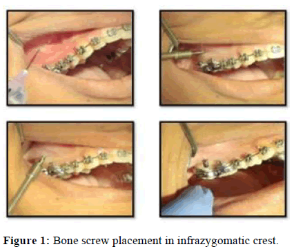
The two posterior IZ screws (Dentos, size 2 mm × 12 mm) were inserted between the first and second molar and 2 mm above the mucogingival junction in the alveolar mucosa. Initially the bone screw was directed at 90° to the Occlusal Plane (OP). After making a initial notch in the bone, the driver was angulated at 55° to 70° toward the teeth. The bone screw was inserted in till only the head of the screw was visible outside the alveolar mucosa (Figure 1).
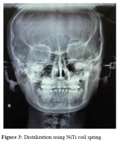
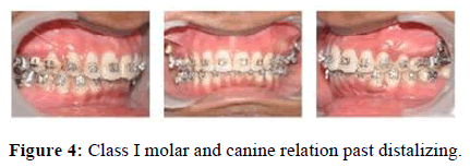
PA view radiographs were taken to check the angulation and the height of insertion of bone screws, also the position of the bone screw in buccal direction for correct vector force application (Figure 2). The screws were loaded immediately with NiTi coil spring and a force of 300 gm per side from mini-implants was applied in the anterior region to crimpable hooks (9 mm) placed between lateral incisor and canine (Figure 3). The distalizing force was continued till class I molar and canine relation was attained (Figure 4).
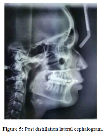
Post-distalization diagnostic aids i.e., Orthopantomogram (OPG), lateral cephalogram (T1), study models intraoral and extra oral photographs, were taken (Figure 5). Lateral cephalograms were traced using Dolphin Imaging software (version 11.7, Dolphin Imaging and Management Solutions, Chatsworth, California). A total of 2 soft tissues, 6 skeletal and 9 dental measurements were made.
Cephalometric parameters
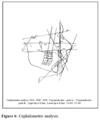
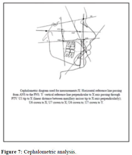
The lateral cephalogram were taken in a centric occlusion at T0 and T1. Sella-Nasion (SN) and Palatal Plane (PP) was taken as horizontal plane and perpendicular to Palatal Plane (PP) through Pterygomaxillary fissure point was taken as vertical plane (Figures 6 and 7).
Skeletal angular measurement
SNA: The anteroposterior relationship of the maxillary basal arch to the anterior cranial base. SNB: The anterior limit of the mandibular base arch in relation to anterior cranial base. ANB: The maxillomandibular relationship to cranial base.
N perpendicular-point A: Sagittal position of maxillary base. N perpendicular-point B: Sagittal position of mandibular base.
Skeletal linear measurement
Ptm-point A: Sagittal position of maxillary base.
Dental angular measurements
SN-U1: Inclination of upper incisor to cranial base.
SN-U6: Inclination of upper first molar to cranial base.
SN-U7: Inclination of upper second molar to cranial base.
Dental linear measurements
In sagittal plane
Ptv-U1: Sagittal position of maxillary central incisor.
Ptv-U6: Sagittal position of maxillary first molar.
Ptv-U7: Sagittal position of maxillary second molar.
In vertical plane
Palatal plane-U6 crown: Vertical position of maxillary first molar.
Palatal plane-U7 crown: Vertical position of maxillary second molar.
Results
Present study evaluated the treatment effects of full arch distalization of maxilla using infrazygomatic crest implants (Dentos). Various parameters were used to evaluate the distalization. Data were analysed using SPSS software v.23 (IBM statistics, Chicago, USA) and Microsoft office 2017. All characteristics were summarized descriptively. The summary statistics of mean ± Standard Deviation (SD) were used for continuous variables. The difference of the means of analysis variables between two time points in same group was tested by paired t-test. P-value less than 0.05, was considered to be statistically significant.
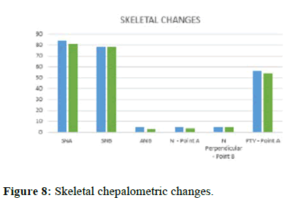
SNA angle observed from pre and post distalization was statistically significant with p>0.05 which indicate that the anteroposterior position of the maxilla with regard to cranial base is significantly changed. There was a statistically significant increase in ANB angle (p>0.05).
Skeletal linear measurement of N⊥-point A showed a statistically significant decrease from pre and post-distalization. Both skeletal angular and skeletal linear measurements of mandible (SNB angle and N ⊥-point B) showed statistically non-significant change post-distalization (Figure 8).
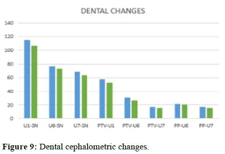
The dental linear measurements of PTV-U1, PTV-U6 and PTV-U7 were found to be statistically significantly. Maxillary incisor retraction of 4.58 mm was seen. Distalization of the maxillary first molar was observed as 4.17 mm along with distal tipping of 3.7°. Distalization of 1.19 mm was observed with the second molar along with increased distal tipping of 4.42° significant retroclination of the maxillary incisor (7.91°) was seen post distalization. Statistically significant amount of intrusion is seen with maxillary first and second molars (1.25 mm and 1.19 mm) (Figure 9).
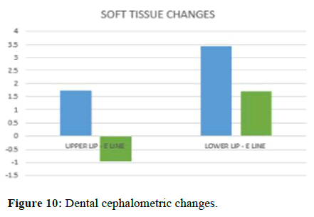
Statistically significant improvement in position of upper and lower lip to E line was observed (p>0.05) (Figure 10 and Table 1).
| Parameters for distalization of maxillary arch | Pre treatment | Post treatment | T2-T1 | P value | Remark | |||
|---|---|---|---|---|---|---|---|---|
| SNA | 84.04 | 0.99 | 81.03 | 1.38 | -2.27 | 0.99 | 0.00012 | S* |
| SNB | 78.56 | 0.79 | 78.66 | 0.96 | 0.1 | 0.5 | 0.5651 | NS |
| ANB | 5.08 | 1.3 | 2.7 | 1.91 | -2.38 | 1.15 | 0.00027 | S* |
| N -⊥Point A | 4.95 | 0.53 | 3.63 | 0.43 | -1.32 | 0.46 | P<0.01* | S* |
| N -⊥Point B | 4.8 | 0.45 | 4.98 | 0.49 | 0.1 | 0.26 | 0.059 | NS |
| PTV-Point A | 56.67 | 1.91 | 54.51 | 2.27 | -2.16 | 0.66 | P<0.01* | S* |
| U1-SN | 115.16 | 3.29 | 107.25 | 2.26 | -7.91 | 3.08 | P<0.01* | S* |
| U6-SN | 77.16 | 1.26 | 73.66 | 1.3 | -3.5 | 1.31 | P<0.01* | S* |
| U7-SN | 68.33 | 1.61 | 63.91 | 1.67 | -4.42 | 0.9 | P<0.01* | S* |
| PTV-U1 | 57.58 | 1.67 | 53 | 0.85 | -4.58 | 1.16 | P<0.01* | S* |
| PTV-U6 | 31 | 1.47 | 26.83 | 1.89 | -4.17 | 1.02 | P<0.01* | S* |
| PTV-U7 | 17.3 | 1.18 | 16.11 | 1.43 | -1.19 | 0.7 | P<0.01* | S* |
| PP-U6 | 22 | 1.34 | 20.75 | 1.48 | -1.25 | 0.62 | P<0.01* | S* |
| PP-U7 | 17.3 | 1.18 | 16.11 | 1.43 | -1.19 | 0.77 | P<0.01* | S* |
| Upper lip-E line | 1.74 | 0.38 | -0.95 | 1.03 | -2.69 | 1.1 | P<0.01* | S* |
| Lower lip-E line | 3.43 | 0.68 | 1.7 | 0.72 | -1.7 | 0.82 | P<0.01* | S* |
Table 1: Conventional cephalometric analysis of the treatment effects of IZC.
Discussion
Orthodontics in its century of existence has a lot of landmarks in its evolution, but very few can match the clinical impact made by recently introduced orthodontic bone screws. The advent of orthodontic bone screws has widened the horizon of orthodontic treatment where borderline extraction and surgical cases can now be treated with non-extraction and non-surgical approaches.
Angle SNA was reduced by 2.27° post total maxillary arch distalization. Comparing with the present study Lee SK et al., observed a clinically non-significant reduction in Angle SNA of 1.07° using modified C palatal plate [8]. Treating class II division 1 malocclusion with gummy smile Shaikh et al. Used IZC bone screw for en-masse distalization and anterior mini-implants for intrusion. 1.1° reduction of SNA angle was observed which was found to be statistically non-significant [9]. In a recent study done by Rosa et al., angle SNA was reduced by 0.45° which was statistically non-significant although in the same study the authors observed statistically significant change in ANB angle [10]. Bechtold et al., performed en-masse distalization using inter-radicular mini-implant and observed no significant skeletal maxillary effects. Doruk et al., in his case report showed a decrease in SNA angle from 79° to 78° [11].
There was a significant decrease in PTV-Point A (2.16 mm) that depicts the correction of dentoalveolar class II pattern. Statistically significant results were observed by Shahani et al., and Shaikh et al., with a similar reduction of 1.89 mm in PTV- Point A.
Statistically significant changes in N ⊥ Point A were observed (1.32 mm) in the present study. On the contrary, Lee SK et al., observed no statistically significant changes in N ⊥ Point A with 0.85 mm. ANB angle showed a statistically significant change of 2.38°. Coincident with the present study Rosa et al., observed a change of 0.75° in ANB angle. SNB angle and PTV–Point B both showed a slight increase of 0.1° which was statistically non-significant. These results were in accordance with studies done by Shaikh et al., Rosa et al., Doruk et al. Molar distalization of 4.17 mm and intrusion of 1.25 mm were observed. The maxillary incisors were retracted 7.91 mm. Wilson Rosa et al., found similar results using infrazygomatic crest implant with molar distalization of 4.7 mm and intrusion of 1.22 mm. Similarly, Wu et al., found distalization of molars (3.5 mm), intrusion of the molars (2.1 mm), and also retraction (4.3 mm) and extrusion (3.8 mm) of the maxillary incisors during a mean treatment time of 8 months using IZC bone screw. Various other case reports showed effective en masse distalization of total maxillary arch [12-15]. A similar magnitude of dental changes was showed by Bechtold et al., who achieved 4.2 mm of distalization and 3.4 mm of retraction of incisors using inter-radicular mini-screw. Comparing the Modified C-Palatal Plate (MCPP) with inter-radicular mini-screws, Lee et al., obtained greater amounts of distalization (4.2 mm) and intrusion (1.6 mm) with less distal tipping of the first molars (2°) and more extrusion of the incisors using MCPP.
Important factor that influenced the results was the relationship between the line of action of the force from bone screw to the crimpable hook and the position of the Center of Resistance (CR) [16]. The IZC bone screws promoted a clockwise rotation of the maxillary occlusal plane because the line of force passed below the CR. But in this present study this force vector was counteracted by long crimpable hooks (9 mm). This prevented rotation of the maxillary occlusal plane. The differences in the amount of tooth displacement seemed to be due to the vertical position of the bone screw and the height of the hooks and variations in the direction of the force vector [17]. The bone screw was implanted in the IZC approximately 14 mm-16 mm above the occlusal plane and the length of the hook was 9 mm.
In the vertical linear measurements, maxillary molar intrusion of -1.25 mm with first molar and -1.19 mm with second molar was observed. Although the intrusion observed in maxillary second molar (-1.19) was less in comparison to study done by Shahani et al., (2.5 mm) and similar to Lee SK et al., (-1.18). This was the result of biomechanics that was applied in the present study where height of crimpable hooks were 9 mm leading to force vector passing through CR of maxillary dentition and achieving en masse bodily distalization of the maxillary arch [17,18]. In the previous studies due to the reduced height of crimpable hooks and vector force passing below the CR, molar intrusion was seen which was not favourable for deep bite corrections.
The upper lip-E line was reduced by 2.69 mm which was statistically significant. Similar results were observed by Shaikh et al., who showed an upper lip retraction of 2.1 mm. Shahani et al., observed a statistically significant change in Upper lip-E line (0.54 mm). This retraction of the upper lip greatly improved the profile of the patient and helped the patient to achieve lip competence. The lower lip-E line reduced by 1.7 mm which was statistically significant, which improved the position of lower lip in relation to the upper anterior.
Conclusions
• Class I molar and class I canine relation was achieved
with total maxillary arch distalization using
infrazygomatic crest bone screw as anchorage.
• Effective molar distalization of -4.17 mm was observed
after en-masse distalization of maxillary arch.
• Significant maxillary incisor retraction of -4.58 mm was
achieved.
Funding
This research did not receive any specific grant from funding
agencies in the public, commercial, or not-for-profit sectors.
Conflict of Interest
None.
References
- Almeida MR, Pereira ALP, Almeida RR, Almeida-Pedrin RR, Silva Filho OG, et al. Prevalence of malocclusion in children aged 7 to 12 years. Dental Press J Orthod. 2011;16:123–131.
- Graber LW, Vanarsdall RL, Vig KWL. Orthodontics: Current Principles and Techniques. 5th edition. Elsevier. Philadelphia. 2012.
- Umale V, Jalgaonkar N, Patil C, Gangadhar MB, Sheth S, et al. Molar distalization-A review. Indian J Orthod Dentofac Res. 2018;4:146-150.
- Cope JB. Temporary anchorage devices in orthodontics: A paradigm shift. Semin Orthod. 2005;11:3-9.
- Melsen B. Mini-implants: Where are we? J Clin Orthod. 2005;39:539.
- Giudice AL, Rustico L, Longo M, Oteri G, Papadopoulos MA, et al. Complications reported with the use of orthodontic miniscrews: A systematic review. Korean J Orthod. 2021;51:199-216.
[Crossref] [Google Scholar] [PubMed]
- Almeida MR. Biomechanics of extra-alveolar mini-implants. Dental Press J Orthod. 2019;24:93-109.
[Crossref] [Google Scholar] [PubMed]
- Lee SK, Abbas NH, Bayome M, Baik UB, Kook YA, et al. A comparison of treatment effects of total arch distalization using modified C-palatal plate vs. buccal miniscrews. Angle Orthod. 2018;88:45-51.
[Crossref] [Google Scholar] [PubMed]
- Shaikh A, Jamdar AF, Galgali SA, Patil S, Patel I, et al. Efficacy of infrazygomatic crest implants for full-arch distalization of maxilla and reduction of gummy smile in class II malocclusion. Dent Pract. 2021;22:1135-1143.
[Crossref] [Google Scholar] [PubMed]
- Rosa WG, de Almeida-Pedrin RR, Oltramari PV, de Castro Conti AC, Poleti TM, et al. Total arch maxillary distalization using infrazygomatic crest miniscrews in the treatment of Class II malocclusion: A prospective study. Angle Orthod. 2023;93:41-8
[Crossref] [Google Scholar] [PubMed]
- Bechtold TE, Kim JW, Choi TH, Park YC, Lee KJ, et al. Distalization pattern of the maxillary arch depending on the number of orthodontic miniscrews. Angle Orthod. 2013;83:266-273.
[Crossref] [Google Scholar] [PubMed]
- Doruk C, Cankaya O, Guvenc I. Non-extraction treatment of skeletal class II adult patient with total maxillary arch distalization. Turk J Orthod. 2015;28:122-128.
- Singh G, Gupta N, Goyal V, Singh R, Izhar A, et al. En-masse distalisation of maxillary arch using TADs (IZC); passive self-ligating appliance vs. clear aligner-A comparative cephalometric study. J Contemp Orthod. 2019;3:11-17.
- Wu X, Liu H, Luo C, Li Y, Ding Y, et al. Three-dimensional evaluation on the effect of maxillary dentition distalization with miniscrews implanted in the infrazygomatic crest. Implant Dent. 2018;27:22-27.
[Crossref] [Google Scholar] [PubMed]
- Yezdani AA, Chatterjee P, Kumar SK, Padmavathy K. Correction of class II skeletal malocclusion with an infra-zygomatic crest bone screw approach. Indian J Public Health Res Dev. 2019;10:2386-2391.
- de Almeida MR. The biomechanics of extra-alveolar TADs in orthodontics. Temporary Anchorage Devices Clinic Orthod. 2020:445-454.
- Sung EH, Kim SJ, Chun YS, Park YC, Yu HS, et al. Distalization pattern of whole maxillary dentition according to force application points. Korean J Orthod. 2015;45:20-28.
[Crossref] [Google Scholar] [PubMed]
- Khan J, Goyal M, Kumar M, Kushwah A, Kaur A, et al. Comparative evaluation of displacement and stress distribution pattern during maxillary arch distalization with infra zygomatic screw-A three dimensional finite element study. Int Orthod. 2021;19:291-300.
[Crossref] [Google Scholar] [PubMed]




 The Annals of Medical and Health Sciences Research is a monthly multidisciplinary medical journal.