Ulcerative Colitis Prone to Delayed Diagnosis in a Nigerian Population: Case Series
2 Departments of Histopathology, Federal Medical Centre, Owerri, Imo State, Nigeria
3 Department of Anatomic Pathology, Federal Staff Hospital Abuja, Nigeria
4 Internal Medicine, Federal Medical Centre, Owerri, Imo State, Nigeria
Citation: Ekwunife CN, Nweke IG, Achusi IB, Ekwunife CU. Ulcerative colitis prone to delayed diagnosis in a Nigerian Population: Case series. Ann Med Health Sci Res 2015;5:311-3.
This open-access article is distributed under the terms of the Creative Commons Attribution Non-Commercial License (CC BY-NC) (http://creativecommons.org/licenses/by-nc/4.0/), which permits reuse, distribution and reproduction of the article, provided that the original work is properly cited and the reuse is restricted to noncommercial purposes. For commercial reuse, contact reprints@pulsus.com
Abstract
Inflammatory bowel disease is an emerging disease burden in the developing world. In Nigeria there is a persisting perception among physicians that it is a very rare disease, and publications on it are sparse. Early manifestations of ulcerative colitis (UC) are therefore likely to be missed at many health institutions. This publication aims to contribute to the growing literature on UC among Nigerians. We present 3 cases of UC that were diagnosed at very late stages. It took a range of 2–7 years for the diagnosis to be made from onset of symptoms. UC was confirmed in the first patient after bowel resection for massive gastrointestinal haemorrhage. The other two had colonoscopy and biopsy for confirmation. An increased awareness about UC is necessary in Nigerian population, because the condition may be commoner than hitherto thought. Provision of colonoscopy services to a wider population will assist in early discovery of this disease.
Keywords
Delayed diagnosis, Nigeria, Ulcerative colitis
Introduction
Inflammatory bowel disease (IBD) has emerged as a global disease. Temporal trends indicate an increasing incidence and prevalence across various regions of the world.[1] Whereas this increase has largely been attributed to environmental influences consequent on urbanization and industrialization, better diagnostic tools and increased awareness by physicians have been contributory. The disease burden may be plateauing in Western nations, but this is not the case in previously low incidence regions like Eastern Europe, Asia and other developing nations.[2] In Nigeria and indeed Africa, there is still the perception among physicians that ulcerative colitis (UC) is a rare disease. However, the increasing reports from Nigeria of this subtype of IBD does suggest that the few case reports and case series in literature may just be the tip of the iceberg.[3-5] This condition is most likely an under diagnosed problem in our environment. We report a case series of UC seen over an 18 months period at two South-eastern Nigerian centres with active gastrointestinal endoscopy services: Federal Medical Centre Owerri and Carez Clinic Owerri. This will serve as a contribution to the wider body of knowledge about UC in Nigeria.
Case Report
Case 1
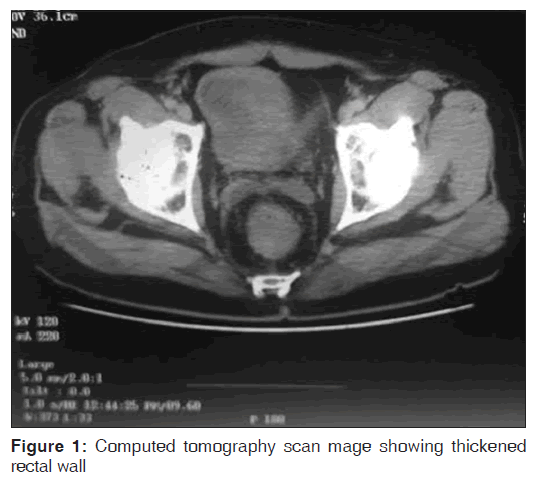
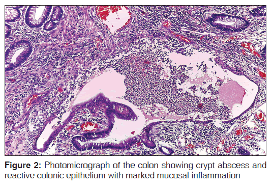
A 64 Nigerian male was referred to our general surgery outpatient clinic on account of 6 months history of progressive weight loss, frequent stooling 5–7 times per day and haematochezia. He had similar bowel complaints 2 years prior to presentation which subsided on unspecified medications. He has had no previous surgical operation. He looked cachetic and anaemic on examination with tenderness on the right upper quadrant of the abdomen. Abdominal computed tomography (CT) done showed thickened bowel which was initially suggested to be a neoplasm of the colon. However, further evaluation of the CT images showed circumferential and diffuse thickening of the rectum as well as the colon; features which are indicative of an inflammatory condition [Figure 1]. On account of the ongoing blood loss he had an emergency laparotomy. There was no tumour found intraoperatively, rather gross inflammation of the entire colon, more pronounced on the descending and sigmoid and transverse colon was noticed. Subtotal colectomy was done. Photomicrograph of the resected bowel segment is as shown [Figure 2]. Postoperatively patient had anastomotic leakage that was managed conservatively. Subsequently, the patient has been maintained on intermittent sulphasalazine for 14 months. During an episode of relapse he was placed on prednisolone but has to be stopped on account of diabetes mellitus. Infliximab was prescribed for him but it has been difficult to access it.
Case 2
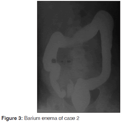
A 30-year-old lady presented with a 7 years history abdominal cramps, 4–9 bowel motions per day that warranted the use of diapers. At the disease onset she was managed in a teaching hospital in neighbouring state where she was admitted for 35 days. During this period she developed widespread suppurative skin lesions. No definitive diagnosis of her condition was made. Subsequently she has been in and out of hospitals on account of febrile illness and abdominal complaints that usually required intravenous fluid and antibiotic resuscitation. A suspicion of UC was made and barium enema requested. It showed loss of haustral markings over the entire length of the colon with evidence of pseudopolyps [Figure 3]. This was confirmed at colonoscopy. She has been on sulphalazine with marked relief of her symptoms.
Case 3
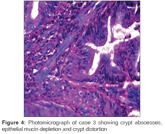
A 41-year-old Nigerian woman presented with a 32 months history of passage of loose stools, 7–8 motions per day, mixed with fresh blood. She occasionally passes blood clots per rectum and has been losing weight. She has received medications at various clinics for diarrhoeal disease without relief. She had erythema nodosum on the dorsum of the left foot. A diagnosis of IBD was suspected and she underwent colonoscopy. Findings included friable mucosal inflammation of the entire rectum and colon as well as a pedunculated 1.5 cm diameter polyp at the descending colon. Histologic sections of the intestinal mucosa tissue fragments showed heavy infiltration of the lamina propria by neutrophils and occasional lymphocytes and plasma cells. Features of crypt abscesses, cryptitis, epithelial damage (mucin depletion) and crypt distortion were also seen [Figure 4]. She was placed on sulphasalazine, which she takes irregularly on account of difficulty in sourcing it. However, stool frequency has decreased to 2–3/day, although it is still bloody.
Discussion
Ulcerative colitis is reportedly very rare in black Africans compared to Western populations.[6] The appreciable data from the disease is mainly from South Africa. This could be attributed to the better health system infrastructure in that country juxtaposed with other sub-Saharan African countries. There are no large patient series from Nigeria despite its population of about 160 million. This becomes more worrisome when neighbouring smaller countries seem to have comparatively larger reported cases. A report from Senegal revealed 32 cases of UC over a 7 years period while from Burkina Faso, Bougouma published 20 cases seen between 1st January 1995 and 21st May 2006.[7,8] Apart from the compounding problems of absence of population based health surveys and adequate medical recording system, the perception that UC is a disease of the Caucasian population points to condition that is potentially under-diagnosed.
Diagnostic delay is a topical issue in the management of IBD in several countries. The Swiss IBD cohort study group reported a median diagnostic delay of 4 months from the onset of symptoms to a diagnosis of UC which is significantly shorter than that for Crohn’s.[9] The alarming nature of bloody diarrhea may be a contributory factor for the earlier diagnosis of UC. However, the delay in the diagnosis of our cases is unduly too long. This may be the reason why all our patients presented with severe disease and associated pancolitis. Patients are likely to self-medicate when they have mild bloody diarrhea as was the case in our first patient. Physicians are also more likely to attribute bloody diarrhea to amoebic colitis, other bacterial infective causes prevalent in the tropics, human immunodeficiency virus and less commonly to colonic neoplasia.[10] Thus it is the severe case that gets to be investigated more comprehensively. Even in this regard options are limited because colonoscopy facilities are still rudimentary in many parts of Nigeria. In our centre, it is barely 1-year. The poor radiologic service in our practice is also being highlighted by this study. The initial CT scan report for Case 1 suggested colonic malignancy as cause of the haemorrhage. It was only in hindsight that another radiologist was able to outline the diffuse thickening of the bowel that is in keeping with an inflammatory process.[11]
As an aid in diagnosis, it needs to be reinforced among our physicians that the skin is the most common extraintestinal organ to be affected in IBD.[12,13] Two of our patients had skin manifestation which heightened our suspicion of UC. A drawback in this regard is that there is a huge dearth of dermatologists in our country.
Treatment of patients with UC has profound challenges in our society. It may be difficult sourcing the basic drug, sulphasalazine. The national drug agency does not seem to have any company that is licensed to import it. Patients occasionally get supplies from neighbouring countries. In order to access infliximab a special application had to be made to the same agency. Ultimately this makes costs prohibitive for patients.
We believe that establishing a national database on UC is pertinent at the moment; as the disease condition becomes increasingly diagnosed and reported in our environment. This will make some sense from the various cases reported from some various health institutions. It will also provide the opportunity for timely and indigenous research on a condition that will likely compound the various health challenges in a sub-Saharan African environment.
REFERENCES
- Molodecky NA, Soon IS, Rabi DM, Ghali WA, Ferris M, Chernoff G, et al. Increasing incidence and prevalence of the inflammatory bowel diseases with time, based on systematic review. Gastroenterology 2012;142:46-54.e42.
- M’Koma AE. Inflammatory bowel disease: An expanding global health problem. Clin Med Insights Gastroenterol 2013;6:33-47.
- Alatise OI, Otegbayo JA, Nwosu MN, Lawal OO, Ola SO, Anyanwu SN, et al. Characteristics of inflammatory bowel disease in three tertiary health centers in southern Nigeria. West Afr J Med 2012;31:28-33.
- Ukwenya AY, Ahmed A, Odigie VI, Mohammed A. Inflammatory bowel disease in Nigerians: Still a rare diagnosis? Ann Afr Med 2011;10:175-9.
- Senbanjo IO, Oshikoya KA, Onyekwere CA, Abdulkareem FB, Njokanma OF. Ulcerative colitis in a Nigerian girl: A case report. BMC Res Notes 2012;5:564.
- Segal I, Tim LO, Hamilton DG, Walker AR. The rarity of ulcerative colitis in South African blacks. Am J Gastroenterol 1980;74:332-6.
- Bougouma A, Sombié R, Ido-Da TR, Zoure N, Darankoum D, Napon-Zong D, et al. Ulcerative colitis in black patients. From 20 cases observed in Burkina Faso. JAfr Hépatol Gastroentérol 2010;4:147-51.
- Diouf ML, Dia D, Thioubou A, Bassène ML, Mbengue M. Prevalence of ulcerative colitis in the digestive endoscopy unit of Aristide-Le-Dantec Hospital of Dakar. J Afr Hépatol Gastroentérol 2010;4:97-102.
- Vavricka SR, Spigaglia SM, Rogler G, Pittet V, Michetti P, Felley C, et al. Systematic evaluation of risk factors for diagnostic delay in inflammatory bowel disease. Inflamm Bowel Dis 2012;18:496-505.
- Nwokediuko S, Bojuwoye B, Ozumba UC, Ozoh G. Peculiarities of chronic diarrhoea in Enugu, Southeastern Nigeria. J Health Sci 2002;48:435-40.
- Fernandes T, Oliveira MI, Castro R, Araújo B, Viamonte B, Cunha R. Bowel wall thickening at CT: Simplifying the diagnosis. Insights Imaging 2014;5:195-208.
- Huang BL, Chandra S, Shih DQ. Skin manifestations of inflammatory bowel disease. Front Physiol 2012;3:13.
- Marzano AV, Borghi A, Stadnicki A, Crosti C, Cugno M. Cutaneous manifestations in patients with inflammatory bowel diseases: Pathophysiology, clinical features, and therapy. Inflamm Bowel Dis 2014;20:213-27.


 The Annals of Medical and Health Sciences Research is a monthly multidisciplinary medical journal.