Unusual Unilateral Multiple Muscular Variations ofBack of Thigh
- *Corresponding Author:
- Mr. Kosuri Kalyan Chakravarthi
Department of Anatomy, Santhiram Medical College, NH-18, Nandyal- 518 501, Kurnool District, Andhra Pradesh, India.
E-mail: kalyankosuric@gmail.com
Citation: Chakravarthi KK. Unusual unilateral multiple muscular variations of back of thigh. Ann Med Health Sci Res 2013;3:S1-2.
Abstract
During routine cadaveric dissection for the undergraduate students in the Department of Anatomy, we noted multiple muscular variations in a middle‑aged male cadaver. All the variations were seen at the back of thigh (flexor compartment) of right lower limb. An anomalous muscle of 17 cm length with average width of 1.5 cm. was inserted to the semitendinosus, a third head of biceps femoris of 6.5 cm length with an average width of 3.5 cm was inserted to the semitendinosus and ununited short and long heads of biceps femoris, both heads were inserted to the head of fibula. To the best of our knowledge, such muscular variations have not been reported in the recent medical literature. A comprehensive knowledge of such rare anatomical variations will be important for surgeons and Traumatologists as this might cause compression of the sciatic nerve.
Keywords
Biceps femoris, Sciatic nerve, Semitendinosus
Introduction
Semitendinosus and biceps femoris belong to the hamstring muscle group in the back of the thigh. Biceps femoris has a long head and short head. Long head belongs to the hamstring part which arises from the lower and inner impression on the back part of the Ischial tuberosity and short head arises from the lateral lip of the linea aspera and from the upper two thirds of lateral supracondylar line of femur. The conjoint tendon of the two heads insert into the lateral side of the head of the fibula and by a small slip into the lateral condyle of the tibia. Semitendinosus arises along with the long head of biceps femoris from the lower and inner impression on the back part of the Ischial tuberosity. Semitendinosus and long head of biceps femoris are innervated by tibial component of the sciatic nerve.
Variations in the hamstring muscle are not common. Knowledge of the presence of muscles variations as well as the location of compression is useful in determining the pathology and appropriate treatment for compressive neuropathies.[1]
Case Report
During routine dissection for the undergraduate students in the Department of Anatomy, Santhiram Medical College, Nandyal, of a middle aged male cadaver, the following multiple muscular variations were observed at the back of thigh of right lower limb.
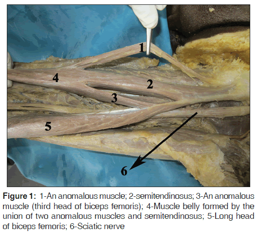
•An anomalous muscle was noted to take from the lower lateral triangular area of ischial tuberosity and adjoing ischiopubic ramus, merging with the semitendinosus. The total length of the muscle from the most proximal point to the most distal point was 17 cm and the average width was 1.5 cm. The muscle received a branch from the tibial nerve on its deep aspect [Figure 1].
• Another anomalous muscle was also seen to pick origin from the long head of biceps femoris and merged with the semitendinosus. The total length of this muscle from the most proximal point to the most distal point was 6.5 cm and the average width was 3.5 cm [Figure 1].
• Ununited short and long heads of biceps femoris, both heads were inserted to the head of the fibula [Figure 2].
Discussion
Proper knowledge of muscular variations is essential not only for anatomists, but also for surgeons. Such muscular variations may lead to error in both diagnosis and treatment. The hamstring muscles [semimembranosus, semitendinosus, and long head of biceps femoris and ischial fibers of the adductor magnus] take origin from the ischial tuberosity and are supplied by tibial component of the sciatic nerve.
Semitendinosus and semimembranosus are considered as true hamstrings. Whereas, the anomalous additional muscle found in this case was aroused from the lateral triangle area of ischial tuberosity (ham string part) and adjoing ischiopubic ramus (non hamstring part), which merged with the semitendinosus. It was innervated by a tibial component of the sciatic nerve. Thus, this additional muscle may be considered as additional head of semitendinosus. Such variations may alter the biomechanics of the muscle.
A search of the literature revealed that an anomalous muscle originated from the long head of the biceps femoris inserted into the tendo calcaneus or sural fascia have been described.[2‑5] Turner et al. reported an anomalous muscle which originated from the long head of the biceps femoris and linea aspera and inserted into the deep surface of the fascia at the superior angle of the popliteal fossa.[6] An anomalous muscle in the popliteal fossa as a muscle arising by 2 thin tendinous slips, one from biceps and the other from semitendinosus and inserting into the tendo calcaneus or at the junction of the two heads of the gastrocnemius muscle has been reported.[7‑9] Where as in this case an anomalous muscle originated from the long head of the biceps femoris merged with semitendinosus has not been reported in modern literature. It may be consider as a third head of biceps femoris. Such anomalous muscle connecting the long head of the biceps femoris to the semitendinosus will certainly improve the strength of the medial rotation in semi‑flexed knee. The variations of such muscles, especially accessory muscles or heads may simulate soft‑tissue tumors and can result in nerve compressions, because of their close relationship to the sciatic nerve. Hence, the clinician must be aware constantly of such possibilities, although preoperative diagnosis may be difficult.
Long head of biceps femoris crosses the middle third of back of thigh from medial to lateral to meet the short head. The conjoint tendon of the two heads inserted to the head of the fibula. Ununited short and long heads of biceps femoris found in this case may contribute to common fibular nerve compression close to the head of the fibula thereby such variation can cause the foot drop.
Conclusion
Failure of muscle primordia to disappear during embryologic development may account for the presence of the accessory or additional muscles reported in this case. Such variations of muscles may simulate soft‑tissue tumors and can result in nerve compressions, because of their close relationship to the sciatic nerve. Hence, awareness of these variations is necessary to avoid complications during radiodiagnostic procedures or surgeries.
Source of Support: Nil.
Conflict of Interest: None declared.
References
- Lahey MD, Aulicino PL. Anomalous muscles associated with compression neuropathies. Orthop Rev 1986; 15: 199-208.
- Halliburton WD. Remarkable abnormality of the musculus biceps flexor cruris. J Anat Physiol 1881; 15: 296-9.
- Turner W. Absence of extensor carpi ulnaris and presense of an accessory sural muscle. J Anat Physiol 1884-5; 19: 333-4.
- Seema SR, Balakrishna. Tensor fascia suralis. J Anat Soc India 2001; 51:130.
- Schaeffer JP. One two muscles anaomalies of the lower extremity. Anat Record1918; 7:1-7.
- Turner W. Muscular system. J Anat Physiol. 1872; 5:441.
- Somayaji SN, Vincent Airy. An anomalous muscle in the region of the popliteal fossa: Case report. J Anat 1998; 192:307-8.
- Montet X, Sandoz A, Mauget D, Martinoli C, Bianchi S. Sonographic and MRI appearance of tensor fasciae suralis muscle, an uncommon cause of popliteal swelling. Skeletal Radiol 2002; 31:536-8.
- Kumar GR, Bhagwat SS. An anomalous muscle in the region of the popliteal fossa: Acase report. J Anat Soc India 2006; 55:65-8.


 The Annals of Medical and Health Sciences Research is a monthly multidisciplinary medical journal.