Accidental Hanging causing complete Laryngotracheal separation: Case Report
2 Department of Medicine, Padhar Hospital, Betul, Madhya Pradesh, India
3 Department of Medicine, Jawaharlal Nehru Medical College, Datta Meghe Institute of Medical Sciences Sawangi (Meghe), Wardha, India
Published: 30-Jul-2021
Citation: Maldhure S, et al. Accidental Hanging Causing Complete Laryngotracheal Separation: Case Report. Ann Med Health Sci Res. 2021;11:18-20.
This open-access article is distributed under the terms of the Creative Commons Attribution Non-Commercial License (CC BY-NC) (http://creativecommons.org/licenses/by-nc/4.0/), which permits reuse, distribution and reproduction of the article, provided that the original work is properly cited and the reuse is restricted to noncommercial purposes. For commercial reuse, contact reprints@pulsus.com
Abstract
Laryngotracheal separation and hanging are both rare. The incidence of laryngotracheal separation is only 0.03% of patients coming to accident and emergency. Similarly, even less literature is available on management of such cases. They are also found to have multiple associated ailments like acute respiratory distress syndrome and vocal cord paralysis, which need to address. Rapid diagnosis and prompt management of airway can save lives. Here, we present a case of accidental hanging with laryngotracheal separation.
Keywords
Laryngotracheal separation; Hanging; Subglottic stenosis; Vocal cord palsy
Introduction
Laryngotracheal separation is a rare finding in patients with acute trauma of neck whether penetrating or blunt trauma. [1] Accidental hanging whether it may be due to automobile accidents or cloth line injury, being uncommon, it makes significant laryngeal injury even more rare. Laryngotracheal trauma patients besides injury to laryngo- tracheal framework, also sustain some degree of insult to recurrent laryngeal nerves causing temporary or permanent paresis or palsy of the vocal cords. [2] Hanging patients are also at risk of developing acute respiratory distress syndrome, specially the incidence being more in young patients with lower GCS. [3] This case report highlights multiple problems associated with accidental hanging, especially laryngotracheal separation.
Case Report
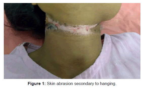
When received, she complained of breathing difficulty, chest pain, vomiting, and pain in neck and hemoptysis. On examination her GCS was 15/15 with signs of respiratory distress, pupils were equal and reacting to light. She was a febrile, pulse-66/ min, BP-140/80 mmhg. Neck had about 10 cm long abrasion at the level of thyroid cartilage with significant swelling (surgical emphysema) of entire neck. Respiratory system showed coarse crepts. Since in respiratory distress with dropping oxygen saturation, she was immediately intubated and started on mechanical ventilation along with nasogastric tube, antibiotics, steroids, bronchodilators, and intermittent nor adrenalin. On compromised fibreopticnaso laryngopharyngoscopy because of intubation, we could see her vocal cords were edematous, their movements could not be appreciated, there was clotted blood in subglottic area and rest of larynx was normal. CT scan neck showed suspicion of laryngotracheal separation below the level of cricoid cartilage with absence of mucosal covering for a length of about 2 cm-3 cm [Figures 1-4].
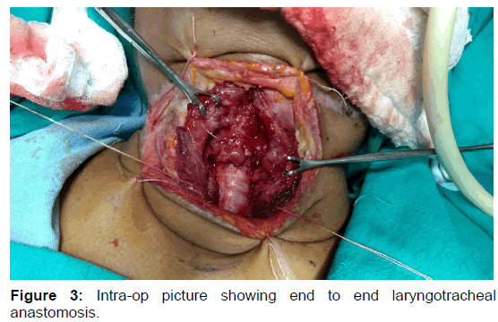
She was taken for emergency neck exploration under general anaesthesia. The skin had abrasion mark at the level of cricoids. The strap muscles were intact but unhealthy looking. There was complete seperation of cricotracheal joint. Esophagus was normal. Trachea along with thyroid gland was lying at the level of clavicle. Inner circumferencial mucosa of cricoid was avulsed and pulled off with trachea. Granulation tissue and mucosa were intervening between cricoid and trachea. Tracheostomy was done in third tracheal ring to secure the airway. Endotracheal tube removed. Cricotracheal anastomosis done with 2 ‘0’ prolene. Portex no.6 endotracheal tube was used as stent. Neck closed in layers. Patient was extubated after 24 hours. NG tube removed on 5th post op day. Neck sutures removed after 8 days. Stent removed after 14 days. Tracheostomy tube changed to metal and care was taught.
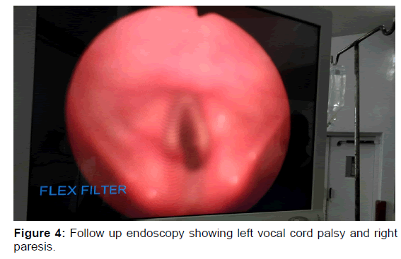
On follow up fibreopticscopy, she was found to have 50 percent subglottic stenosis with left vocal cord palsy and right vocal cord paresis. She was advised laser assisted dilatation of subglottis.
Discussion
Injury to the laryngotracheal framework due to blunt or penetrating trauma is less common due to its protective anatomical arrangement with mandible above and sternum below & bulky sternocleidomastoid muscles on either side. Thus, making laryngotracheal separation even more rarer. The incidence being reported as only 0.03% of patients coming to accident and emergency department. [4]
Our patient with accidental hanging presented as acute respiratory distress syndrome to accident and emergency centre, she was immediately intubated and started on mechanical ventilation with supportive care. ARDS could have been secondary to aspiration or neurogenic pulmonary edema. [3] It is difficult to say here whether intubation is justified or not because the type and amount of damage caused by the event was not assessed completely as described by Schaefer et al. [5] The airway integrity was not confirmed clearly. But the emergency of situation cannot be ignored, and according to availibity of resources at hand patient was intubated. In such circumstances, it is possible to displace the distal end of severed airway, making resusitation of patient even more difficult.
On futher evaluation she was found to have complete laryngotracheal separation and bilateral recurrent laryngeal nerve injuries. She probably sustained this injury because of the sudden flexion and extension of the neck caused by loose cloth draped around her neck got stuck to a pole when she suddenly slipped. The chances of laryngotracheal separation by the force of sudden flexion and extension are more than by direct blows which more often cause fracture of cartilaginous framework. [6] Flexible fibreoptic laryngoscopy and CT scan imaging helped to establish the diagnosis. Cricotracheal anastomosis was done after securing airway, and stent was placed. Placing a stent also has been a controversial issue, since it has complications like granulation, crusting, etc. But here authors are of view that in our case it is the stent which prevented the subglottis from complete stenosis and secured 50% airway.
On follow up, she was found to have left vocal cord palsy and right vocal cord paresis. The vocal cords were not observed during surgery. It is difficult to figure out reason for vocal cord injury, whether it is due to hanging, tracheal intubation or surgical procedure. A previous report indicated that management options for vocal cord paralysis vary depending on the underlying etiology. [7] Kunii et al. reports, reversal of vocal cord paralysis without surgical intervention attributing it to edema of laryngeal muscles following neck injury. [8] It is very difficult to explore for recurrent laryngeal nerves in a case of injury to the laryngotracheal framework. However, in cases where nerves are identified and its severed ends are found, they can be end to end anastomosed or can be repaired using nerve graft if they are separated by more than 5 millimeters. [9] Studies on related aspects of laryngotracheal structures were reviewed. [10-15]
Conclusion
There is scarcity of literature on laryngotracheal separation due to less incidence, could be due to immediate death of patients or cases going undiagnosed. High index of suspicion for laryngotracheal separation and vocal cord paralysis is thus very important in cases of trauma to neck or hanging. Rapid diagnosis and prompt management is the key towards saving patient. Airway should be addressed first, once secured acute respiratory distress syndrome should be ruled out followed by surgical management of laryngotracheal separation. Recurrent laryngeal nerves should be looked for and appropriate intervention should be done. Finally, all preventive measures should be taken to avoid such accidents and save precious lives.
References
- Plocienniczak MJ, Finlay S, Noordzij JP, Cohen MB. Case report: Complete laryngotracheal separation sustained from a knife wound. Otolaryngology Case Reports. 2019;11:1-3.
- Latoo M, Lateef M, Nawaz I, Ali I. Bilateral recurrent laryngeal nerve palsy following blunt neck trauma. Indian J Otolaryngol Head Neck Surg. 2007;59:298-299.
- Mansoor S, Afshar M, Barrett M, Smith GS, Barr EA, Matthew E, et al. Acute respiratory distress syndrome and outcomes after near hanging. Am J Emerg Med. 2015;33:359-362.
- Gussack GS, Jurkovich GJ, Luterman A. Laryngotracheal trauma: A protocol approach to a rare injury. Laryngoscope. 1986;96:660-665.
- Schaefer SD. Management of acute blunt and penetrating external laryngeal trauma. Laryngoscope. 2014;124:233-244.
- Mathisen DJ, Grillo H. Laryngotracheal trauma. Ann Thorac Surg. 1987;43:254-262.
- Maisel RH, Ogura JH. Evaluation and treatment of vocal cord paralysis. Laryngoscope. 1974;84:302-316.
- Kunii M, Ishida K,Ojima M,Sogabe T, Shimono K, Tanaka T, et al. Bilateral vocal cord paralysis in a hanging survivor: A case report. Acute Med Surg. 2020;7:1-4.
- Lynch J, Parameswaran R. Management of unilateral recurrent laryngeal nerve injury after thyroid surgery: A review. Head Neck. 2017;39:1470-1478.
- Ansari I, Bagga CS, Garikapathi A, Naik S, Kumar S. Takot subocardiomyopathy: Case Report after Attempted Suicidal Partial Hanging. Medi Sci. 2020;24:2435-2438.
- Nameirakpam C, Hijam B, Ninave S, Singh ST, Keisham U, Heisnam I. A double blind comparative study of clonidine and fentanyl to see the haemodynamic response during laryngoscopy and intubation. JEMDS. 2014;3:10015-10025.
- Golhar S. Tracheooesophageal puncture voice: An indian perspective. Indian J Otolaryngol Head Neck Surg.2014;62:408-414.
- BholaN, Jadhav A, Borle R, Khemka G, Ajani AA. Awake endotracheal retrograde intubation in restricted mouth opening: A `J’-tipped guide wire technique: A Retrospective Study. Oral Maxillofac Surg. 2014;18:393-396.
- Chandrakant P, Patil R, Biswas J, Nahata V. Rhinoscleroma with laryngotracheal involvement, causing diagnostic dilemma. JEMDS. 2013;3:395-399.
- TidkeAS, Borle RM, Madan RS, Bhola ND, Jadhav AA, Bhoyar AG. Transmylohoid submental endotracheal intubation in pan- facial trauma: A paradigm shift in airway management with prospective study of 35 cases. Indian J Otolaryngol Head Neck Surg. 2013;65:255-259.


 The Annals of Medical and Health Sciences Research is a monthly multidisciplinary medical journal.