Dental Caries Status in Children with and without Cleft Lip and Palate: A Case Control Study
2 Department of Pedodontics, Saveetha Dental College And Hospitals, Saveetha Institute Of Medical And Technical Sciences, Saveetha University, Chennai, India, Email: vigneshr.sdc@saveetha.com
Received: 02-Sep-2021 Accepted Date: Sep 09, 2021 ; Published: 23-Jul-2021
This open-access article is distributed under the terms of the Creative Commons Attribution Non-Commercial License (CC BY-NC) (http://creativecommons.org/licenses/by-nc/4.0/), which permits reuse, distribution and reproduction of the article, provided that the original work is properly cited and the reuse is restricted to noncommercial purposes. For commercial reuse, contact reprints@pulsus.com
Abstract
Prevalence of dental caries in children with cleft lip and palate was shown to be high compared to any other oral complications. The prevalence of associated systemic disease and those are defected postnatally is 25% among children with cleft. The number of factors such as literacy level of their parents, socioeconomic status and occupation of parents also contributes to the poor oral hygiene status of those children. To determine the dental caries status in children with and without cleft lip and palate. The purpose of the study to analyse the dental caries status in children with and without cleft lip and palate. A study was carried out by collecting data by reviewing patients data and analysing the data of 86000 patients between June 2019 and March 2020 at the private dental institute. The sample size that was taken included 5 children with cleft lip and palate (case group) and 5 children without cleft lip and palate (control group), who came to the private dental institute for consultation. The dental caries status were evaluated using DMFT/dmft score and was compared between the groups. Data was statistically analysed using SPSS 2.0, Mann-Whitney U Test was conducted. Result was recorded. The results showed that children with cleft lip and palate showed a lesser DMFT score compared to children without cleft lip and palate, statistically significant (Mann-Whitney U-test; p=0.034). Therefore it was concluded that within the limitation of the current study, children with cleft lip and palate showed lesser incidences of dental caries when compared to children without cleft lip and palate.
Keywords
Dental caries; DMFT score; Prevalence; Cleft; Lip; Palate; Children
Introduction
Dental caries in children has been increasing in many populations and has become a significant health problem in many countries. [1] Early childhood caries is defined as the presence of one or more decayed, missing or filled tooth surfaces in any primary tooth in a child at 71 months age or younger. [2,3] There are few unique characteristics in clinical appearance of dental caries in children such as rapid development of discoloration on the tooth surface, which affects a number of teeth soon after they emerge in the oral cavity. [4] Major etiology which were correlated with early rampant caries, baby bottle caries, milk bottle syndrome and prolonged nursing habit caries. [5] Dental caries in children are known to be multifactorial disease that results from the interaction of factors that include cariogenic microorganisms, exposure to fermentable carbohydrates through in appropriate feeding practices and a range of social variables. [6,7] Dental caries are not only harmful for normal children, an extra care has to be taken for those children with congenital defects such as cleft lip and palate. [8]
This congenital defect is defined as congenital birth defect which is characterised by complete or partial cleft of lip and palate. [9,10] The severity of the cleft may vary from the trace of notching of the upper lip to a complete non fusion of the lip, the primary palate and the secondary palate. [11,12] Cleft lip and palate anomaly constitute nearly one- third of all congenital malformations of the craniofacial region with an average worldwide incidence of 1 in 700. [13] Its incidence in Asian population is reported to be around two per thousand live births or higher. In India, even though a national epidemiological data is not available, many studies from different parts of the country have reported a variation in the incidence of cleft lip and palate in North India to be 1.4 per 100 births. Mossey et al. estimated from various multicentric studies across India that the incidence of cleft lip and palate in this country ranges from around 0.93-1.3 for cleft lip and palate. [14]
Based on rough estimation, it is suggested that approximately 35000 newborns with cleft lip and palate are added every year into the India population. [15] With many cleft patients having less than optimum care in a not well organized setup, the cumulative burden of children affected with this birth effect is huge. [16] Although India has a large and extended network of medical facilities, interdisciplinary cleft care is provided only in few hospitals. [17] In addition, prevalence of dental caries in children with cleft lip and palate was shown to be high compared to any other oral complications. [18] The prevalence of associated systemic disease and those are defected postnatally is 25% among children with cleft. [19]
However, majority of previous studies have been reported that children with this orofacial cleft have high frequency of dental caries and they lead a poor quality of life. [20,21] Previously our team has a rich experience in working on various research projects across multiple disciplines. [22–36] Now the growing trend in this area motivated us to pursue this project.
The number of factors such as literacy level of their parents, socioeconomic status and occupation of parents also contributes to the poor oral hygiene status of those children. [37] Therefore, present study was conducted to evaluate the prevalence of dental caries in children with cleft lip and palate.
Patients and Methods
This retrospective study was conducted under a hospital based university setting. Ethical approval for this study was granted by the institute’s ethical committee (ethical approval number: SDC/SIHEC/2020/DIASDATA/0619-0320). Consent to use treatment records for research purposes were obtained from patient/ guardian at the time of patient entry into the university for dental needs. The retrospective data were collected by obtaining and analysing the 89000 dental case records of the university from June 2019 to March 2020. The sample size that was taken are 10 children which includes 5 children with cleft lip and palate (case group) and 5 children without cleft lip and palate (control group), who came to the private dental institute for consultation. The inclusion criteria for the current study were children with cleft lip and palate, children between the age of 3-17 years age, complete photographic and written records regarding the complete intra-oral examination of the patient. Age and gender matched controls i.e., children without cleft palate, were taken according to the relevant cases obtained from the inclusion criteria. The exclusion criteria were incomplete and censored dental records, absence of photographic evidence and children below the age of 3 were excluded.
DMFT/deft scores were evaluated for each of the children from both the groups. The selected case and control group were examined by three people; one reviewer, one guide and one researcher. The patients’ case sheets were reviewed thoroughly. Cross checking of data including digital entry and intra oral photographs was done by an additional reviewer and as a measure to minimise sampling bias, samples for the group were picked by simple random sampling. Digital entry of clinical examinations and intra oral photographs of selected subjects were assessed and this included the assessment of tooth decay as mentioned before by the examiner based on intraoral photographs and clinical examination data for each tooth. The examiner was trained to add data of dental caries as present or absent for both case and control group by tabulation using Microsoft Excel software. The mentioned data were coded and transferred into SPSS PC version 2.0 (IBM 2019) software for statistical analysis. A correlation test, Mann-Whitney U Test was done between children with cleft lip and palate (case) and children without cleft lip and palate (control). The results were recorded. The difference was considered statistically significant as the p value was less than 0.05 (p<0.05).
Results and Discussion
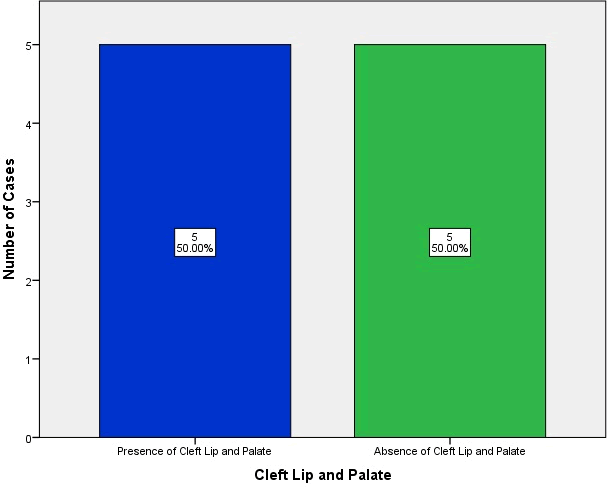
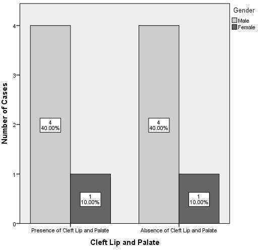
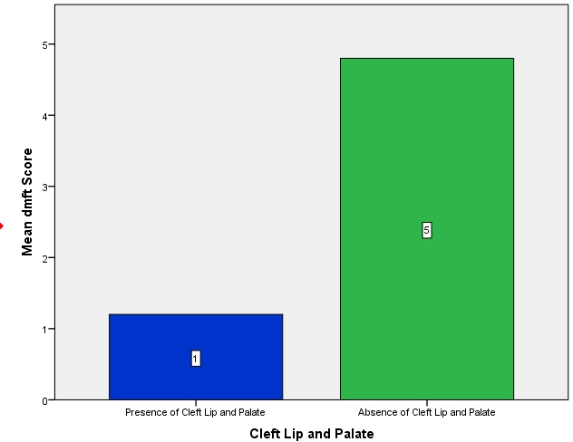
Having cleft lip and palate (case group) and five patients (50%) without cleft lip and palate (control group). It showed the equal distribution of cases in this study [Figure 1]. Among the 10 patients in both the groups, four were males (80%) and one was female (20%), respectively in both groups [Figure 2]. The mean DMFT/deft score was 1.2 for the case group (children with cleft lip and palate) and 4.8 for the control group (children without cleft lip and palate), which was statistically significant (p-value=0.034) [Figure 3]. The Mean DMFT score of children without cleft lip and palate was higher in males [5] when compared to females, [3] but was similar in both males and females with cleft lip and palate. [1] This difference was not statistically significant. (pvalue=0.54) [Figure 4].
Figure 1: The graph shows case distribution in case group (children with cleft lip and palate) and control group (children without cleft lip and palate). (X axis: Presence or absence of cleft lip and palate; Y axis: Number of cases, Blue: Presence of cleft lip and palate in children; Green: Absence of cleft lip and palate in children). Notice the equal distribution of cases for both the groups.
Figure 2:The graph shows gender distribution in case group (children with cleft lip and palate) and control group (children without cleft lip and palate). (X axis: Presence or absence of cleft lip and palate; Y axis: number of cases; Darker Grey: Females; Lighter Grey: Males). Notice the equal distribution of gender for both the groups.
Figure 3:The graph bar portrays the dmft index score distribution for case (with cleft lip and palate) and for control (without cleft lip and palate) groups. (X-axis: Present or absent of cleft lip and palate; Y-axis: Mean dmft score; Blue: Presence of cleft lip and palate in children; Green: Absence of cleft lip and palate in children). Lower dmft score noted in children with cleft lip and palate when compared to children without cleft lip and palate. (Mann-Whitney Test; p=0.034-significant).
Figure 4: The graph bar depicts the comparison of DMFT index score against the gender of children with cleft lip and palate and children without cleft lip and palate. (X-axis: Gender of the patient; Y-axis: Mean dmft score; Blue: Presence of cleft lip and palate in children; Green: Absence of cleft lip and palate in children). The Mean DMFT score of children without cleft lip and palate was higher in males (5) when compared to females (3), which was not statistically significant. (Mann Whitney test; p=0.54-not significant).
A similar study by Rubina et al. [38] revealed that dmft scores among non-cleft children were significantly more (p<0.01) as compared to children with cleft lip and/or palate. This result was similar to the parent study. In another study by Ali et al. [39] showed that children with cleft lip and/or palate had the highest prevalence of dental caries compared to children without cleft lip and/or palate. They have concluded that children with cleft lip and/or palate have higher dmft score due to their diet which is conducive for the development of mild or moderate dental caries. This study was contradictory to the parent study.
In addition, a study conducted by Lucas et al. [40] showed that they found no significant difference in isolated frequency of Streptococcus Mutans in twenty five children aged 3-15 years old children with cleft lip and/or palate and their age matched the control, while Bokhou et al. [41] conducted a study on 18 month old children with cleft lip and/ or palate revealed that they were highly prone to dental caries as S. mutans could be isolated from the saliva of 85% of the children. As a summary of the present study, children with cleft lip and palate had less DMFT/deft score compared to children without cleft lip and palate. The results showed significant difference in the dental caries status for both the groups (p<0.05). The consensus for this study was disagreed as the study is limited to certain factors.
The advantages of the study were that this was a case-control study with age and gender matched control to provide better results and high internal validity. The limitations of the study were smaller sample size (10 childrens only), geographic limitations, unicentric study (based on only one private dental institute), and only one ethnic group was evaluated. Moreover, the possible reasons that can be analysed for caries prone patients are due to poor oral hygiene and their high caries risk diet, which were not considered in this study. For the future scope, the public/parents should be provided with awareness and assessment on the baseline of oral health in children with or without cleft lip and palate. An aggressive approach should be given to the children and parents by practitioners to prevent oral disease and optimize the clinical outcome for patients with or without cleft lip and palate. Our institution is passionate about high quality evidence based research and has excelled in various fields. [42–48] We hope this study adds to this rich legacy.
Conclusion
Within the limitations of the current study, children with cleft lip and palate had lower incidence of dental caries when compared to children without cleft lip and palate.
Acknowledgement
The authors of this study acknowledge the institute, for their help towards collecting all the patient case records and other data in relevance to the current study.
Author Contributions
• Design-KausalyahKrisna Malay, VigneshRavindran
• Intellectual content-VigneshRavindran
• Data collection-KausalyahKrisna Malay
• Data analysis-VigneshRavindran, Jayanth Kumar
• Manuscript writing-KausalyahKrisna Malay
• Manuscript editing-VigneshRavindran, Jayanth Kumar
Conflict of Interest
The authors declare that there were no conflicts of interest.
REFERENCES
- Jeevanandan G. Kedo-S paediatric rotary files for root canal preparation in primary teeth–Case report. J Clin Diagn. 2017;11:ZR03-ZR05.
- Govindaraju L, Jeevanandan G, Subramanian EMG. Comparison of quality of obturation and instrumentation time using hand files and two rotary file systems in primary molars: A single-blinded randomized controlled trial. Eur J Dent. 2017;11:376–9.
- Govindaraju L, Jeevanandan G, Subramanian EMG. Knowledge and practice of rotary instrumentation in primary teeth among Indian dentists: A questionnaire survey. Int J Oral Health Dent. 2017;9:45.
- Chopra A, Lakhanpal M, Rao NC, Gupta N, Vashisth S. Oral health in 4-6 years children with cleft lip/palate: a case control study. N Am J Med Sci. 2014;6:266–9.
- Somasundaram S. Fluoride content of bottled drinking water in Chennai, Tamilnadu. J Clin Diagn. 2015;9:ZC32-ZC34.
- Jeevanandan G, Govindaraju L. Clinical comparison of Kedo-S paediatric rotary files vs manual instrumentation for root canal preparation in primary molars: a double blinded randomised clinical trial. Eur Arch Paediatr Dent. 2018;19:273–8.
- Govindaraju L, Jeevanandan G, Subramanian E. Clinical evaluation of quality of obturation and instrumentation time using two modified rotary file systems with manual instrumentation in primary teeth. J Clin Diagn Res. 2017;11:ZC55–8.
- Zhu WC, Xiao J, Liu Y, Wu J, Li JY. Caries experience in individuals with cleft lip and/or palate in China. Cleft Palate Craniofac J. 2010;47:43–7.
- Ravikumar D, Jeevanandan G, Subramanian EMG. Evaluation of knowledge among general dentists in treatment of traumatic injuries in primary teeth: A cross-sectional questionnaire study. Eur J Dent. 2011;11:232–7.
- Panchal V, Jeevanandan G, Subramanian E. Comparison of instrumentation time and obturation quality between hand K-file, H-files, and rotary Kedo-S in root canal treatment of primary teeth: A randomized controlled trial. J Indian Soc Pedod Prev Dent. 2019;37:75–9.
- Christabel SL, Gurunathan D. Prevalence of type of frenal attachment and morphology of frenum in children, Chennai, Tamil Nadu. World J Dent. 2015;6:203-207
- Packiri S, Gurunathan D, Selvarasu K. Management of paediatric oral ranula: A systematic review. J Clin Diagn Res. 2017;11:ZE06–9.
- Al-Dajani M. Comparison of dental caries prevalence in patients with cleft lip and/or palate and their sibling controls. Cleft Palate Craniofacial Journal. 2009;46:529–31.
- Mossey P, Little J. Addressing the challenges of cleft lip and palate research in India. Indian J Plast Surg. 2009;42:S9–18.
- Gurunathan D, Shanmugaavel AK. Dental neglect among children in Chennai. J Indian Soc Pedod Prev Dent. 2016;34:364–9.
- Govindaraju L, Gurunathan D. Effectiveness of chewable tooth brush in children-A prospective clinical study. J Clin Diagn Res. 2017;11:ZC31–4.
- Sundell AL, Nilsson AK, Ullbro C, Twetman S, Marcusson A. Caries prevalence and enamel defects in 5-and 10-year-old children with cleft lip and/or palate: A case-control study. Acta Odontol Scand. 2016;74:90–5.
- Subramanyam D, Gurunathan D, Gaayathri R, Vishnu Priya V. Comparative evaluation of salivary malondialdehyde levels as a marker of lipid peroxidation in early childhood caries. Eur J Dent. 2018;12:67–70.
- Edlund K, Koch G. Effect on caries of daily supervised tooth brushing with sodium monofluorophosphate and sodium fluoride dentifrices after 3 years. Scand J Dent Res. 1977;85:41–5.
- Ramakrishnan M, Bhurki M. Fluoride, fluoridated toothpaste efficacy and its safety in children-review. IJPR. 2018;10:109–14.
- Lakshmanan L, Mani G, Jeevanandan G, Ravindran V, Ganapathi SEM. Assessing the quality of root canal filling and instrumentation time using kedo-s files, reciprocating files and k-files. Braz Dent Sci. 2020;23:7-10.
- Ponnulakshmi R, Shyamaladevi B, Vijayalakshmi P, Selvaraj J. In silico and in vivo analysis to identify the antidiabetic activity of beta sitosterol in adipose tissue of high fat diet and sucrose induced type-2 diabetic experimental rats. Toxicol Mech. 2019;29:276–90.
- Mathew MG, Samuel SR, Soni AJ, Roopa KB. Evaluation of adhesion of Streptococcus mutans, plaque accumulation on zirconia and stainless steel crowns, and surrounding gingival inflammation in primary molars: randomized controlled trial. Clin Oral Investig. 2020;24:3275–80.
- Subramaniam N, Muthukrishnan A. Oral mucositis and microbial colonization in oral cancer patients undergoing radiotherapy and chemotherapy: A prospective analysis in a tertiary care dental hospital. J Investig Clin Dent. 2019;10:e12454.
- Girija ASS, Shankar EM, Larsson M. Could SARS-CoV-2-induced hyperinflammation magnify the severity of coronavirus disease (covid-19) leading to acute respiratory distress syndrome? Front Immunol. 2020;27:1206.
- Dinesh S, Kumaran P, Mohanamurugan S, Vijay R, Singaravelu DL, Vinod A, et al. Influence of wood dust fillers on the mechanical, thermal, water absorption and biodegradation characteristics of jute fiber epoxy composites. J Polym Res. 2020;27.
- Thanikodi S, Singaravelu DK, Devarajan C, Venkatraman V, Rathinavelu V. Teaching learning optimization and neural network for the effective prediction of heat transfer rates in tube heat exchangers. Therm Sci. 2020;24:575–81.
- Murugan MA, Jayaseelan V, Jayabalakrishnan D, Maridurai T, Kumar SS, Ramesh G, et al. Low velocity impact and mechanical behaviour of shot blasted SiC wire-mesh and silane-treated aloevera/hemp/flax-reinforced SiC whisker modified epoxy resin composites. Silicon Chem. 2020;12:1847–56.
- Vadivel JK, Govindarajan M, Somasundaram E, Muthukrishnan A. Mast cell expression in oral lichen planus: A systematic review. J Investig Clin Dent. 2019;10:e12457.
- Chen F, Tang Y, Sun Y, Veeraraghavan VP, Mohan SK, Cui C. 6-shogaol, an active constiuents of ginger prevents UVB radiation mediated inflammation and oxidative stress through modulating NrF2 signaling in human epidermal keratinocytes (HaCaT cells). J Photochem Photobiol B. 2019;197:111518.
- Manickam A, Devarasan E, Manogaran G, Priyan MK, Varatharajan R, Hsu CH, et al. Score level based latent fingerprint enhancement and matching using SIFT feature. Multimed Tools Appl. 2019;78:3065–85.
- Wu F, Zhu J, Li G, Wang J, Veeraraghavan VP, Krishna Mohan S, et al. Biologically synthesized green gold nanoparticles from induce growth-inhibitory effect on melanoma cells (B16). Artif Cells Nanomed Biotechnol. 2019;47:3297–305.
- Ma Y, Karunakaran T, Veeraraghavan VP, Mohan SK, Li S. Sesame inhibits cell proliferation and induces apoptosis through inhibition of STAT-3 translocation in thyroid cancer cell lines (FTC-133). Biotechnol Bioprocess Eng. 2019;24:646–52.
- Ponnanikajamideen M, Rajeshkumar S, Vanaja M, Annadurai G. In vivo type 2 diabetes and wound-healing effects of antioxidant gold nanoparticles synthesized using the insulin plant Chamaecostus cuspidatus in albino rats. Can J Diabetes. 2019;43:82–9.e6.
- Vairavel M, Devaraj E, Shanmugam R. An eco-friendly synthesis of Enterococcus Sp.-mediated gold nanoparticle induces cytotoxicity in human colorectal cancer cells. Environ Sci Pollut Res Int. 2020;27:8166–75.
- Paramasivam A, VijayashreePJ, Raghunandhakumar S. N6-adenosine methylation (m6A): a promising new molecular target in hypertension and cardiovascular diseases. Hypertens Res. 2020;43:153–4.
- Mutarai T, Ritthagol W, Hunsrisakhun J. Factors influencing early childhood caries of cleft lip and/or palate children aged 18 to 36 months in southern Thailand. Cleft Palate Craniofac J. 2008;45:468–72.
- Shashni R, Goyal A, Gauba K, Utreja AK, Ray P, Jena AK. Comparison of risk indicators of dental caries in children with and without cleft lip and palate deformities. Contemp Clin Dent. 2015;6:58–62.
- Ali YA, Chandranee NJ, Wadher BJ, Khan A, Khan ZH. Relationship between caries status, colony forming units (cfu) of Streptococcus mutans and Snyder caries activity test. J Indian Soc Pedod Prev Dent. 1998;16:56–60.
- Lucas VS, Gupta R, Ololade O, Gelbier M, Roberts GJ. Dental health indices and caries associated microflora in children with unilateral cleft lip and palate. Cleft Palate Craniofac J. 2000;37:447–52.
- Bokhout B, van Limbeek J, Kramer GJC, Prahl-Andersen B. Incidence of dental caries in the primary dentition in children with a cleft lip and/or palate. Caries Res.1997;31:8–12.
- VijayashreePJ. In silico validation of the non-antibiotic drugs acetaminophen and ibuprofen as antibacterial agents against red complex pathogens. J Periodontol. 2019;90:1441–8.
- Ezhilarasan D, Apoorva VS, Ashok VN. Syzygium cumini extract induced reactive oxygen species-mediated apoptosis in human oral squamous carcinoma cells. J Oral Pathol Med. 2019;48:115–21.
- Ramesh A, Varghese S, Jayakumar ND, Malaiappan S. Comparative estimation of sulfiredoxin levels between chronic periodontitis and healthy patients-A case-control study. J Periodontol. 2018;89:1241–8.
- Mathew MG, Samuel SR, Soni AJ, Roopa KB. Evaluation of adhesion of Streptococcus mutans, plaque accumulation on zirconia and stainless steel crowns, and surrounding gingival inflammation in primary molars: randomized controlled trial. Clin Oral Investig. 2020;24:3275–3280
- Sridharan G, Ramani P, Patankar S, Vijayaraghavan R. Evaluation of salivary metabolomics in oral leukoplakia and oral squamous cell carcinoma. J Oral Pathol Med. 2019;48:299–306.
- Pc J, Marimuthu T, Devadoss P. Prevalence and measurement of anterior loop of the mandibular canal using CBCT: A cross sectional study. Clin Implant Dent Relat Res. 2018;20:531-534
- Ramadurai N, Gurunathan D, Samuel AV, Subramanian E, Rodrigues SJL. Effectiveness of 2% Articaine as an anesthetic agent in children: randomized controlled trial. Clin Oral Investig. 2019;23:3543–50.


 The Annals of Medical and Health Sciences Research is a monthly multidisciplinary medical journal.