Sialochemistry and Dental Caries in Down’s syndrome: A Gel Electrophoresis
2 Department of Prosthodontics, Lenora Institute of Dental Sciences, Rajahmundary, Andhra Pradesh, India
3 Department of Public Health Dentistry, Lenora Institute of Dental Sciences, Rajahmundary, Andhra Pradesh, India
Citation: Punithavathy R, et al. Sialochemistry and Dental Caries in Down’s syndrome:A Gel Electrophoresis. Ann Med Health Sci Res. 2018;8:70-73
This open-access article is distributed under the terms of the Creative Commons Attribution Non-Commercial License (CC BY-NC) (http://creativecommons.org/licenses/by-nc/4.0/), which permits reuse, distribution and reproduction of the article, provided that the original work is properly cited and the reuse is restricted to noncommercial purposes. For commercial reuse, contact reprints@pulsus.com
Abstract
Background: Scientific clue of awareness, perceptivity to dental caries in the population with Down’s syndrome is hampered and adverse, making it challenging to establish firm conclusions. Wide range of developmental delays and physical disabilities caused by a genetic disease, Down’s syndrome, which is caused when abnormal cell division results in extra genetic material from chromosome 21. This disease has an incidence of 1 in 600-700 live births. One of the most prominent oral manifestations of Down’s syndrome is low incidence of dental caries. Diminished salivary pH and bicarbonate levels. Biochemical alterations in saliva, delayed eruption, less susceptibility to cariogenic environment, shallow fissures of teeth, all contribute to the lower risk of dental caries. Aim: To compare the glycoproteins, proline rich proteins in children with Down’s syndrome and in caries free children. Settings and Design: The study was conducted in 5-18 years old 30 children comprising two groups Group-1: 15 children with Down’s syndrome, Group-2: 15 caries free healthy children. Materials and Methods: Glycoproteins, proline rich proteins by SDS-PAGE were compared among both the groups. Statistical Analysis: Unpaired t-test, chi-square test was used to compare the data between the study and control group. Results: The out cropping shows no statistical difference in the number of PRP bands in either of the groups. Conclusion: The present study results highlight the central axial dominant role played by PRP’s might be the reason in the protection against dental caries and further detection by monoclonal antibody or LC/MS analysis and other assays to support the result at nano level which escort to burnish the potentiality of its use in caries prediction.
Keywords
Down’s syndrome; Salivary pH; Gel electrophoresis; Proline rich proteins
Introduction
Harmony of life lies in the proportional activity of physical and mental magnitude of health coupled with an ideal social environment, but sometimes an impairment or disability might occur that borders, anticipate the achievement of a role that is normal for an individual. Therefeore a mental illness can seriously impair, temporarily or permanently the mental functioning of a person. Nearly 83 million of the world’s population is estimated to be mentally handicapped. [1] One of the pronounced causes of mental retardation is Down’s syndrome.
It is habitually blended with a delay in cognitive ability (MR) and physical growth with a meticulous set of facial characteristics. The incidence of Down’s syndrome is of 1 in 600-700 live births. [2] It is an easily recognized, congenital, autosomal anomaly caused either due to duplication of a bit of chromosome 21 (Trisomy 21) or translocation. With gradual multiplying of maternal age, the incidence of Down’s syndrome increases as well. [3] Apart from variable physical distinguishability and subnormal mentality in Down’s syndrome, there are specific intra oral appearances like congenital oligodontia, delayed eruption of primary and permanent teeth, malocclusion, mouth breathing leading to dry mouth, increased incidence of periodontitis and soft tissue forces resulting in impaired chewing and consequently difficulty in self cleansing of teeth [4-8] which are predisposing factors for caries aetiology.
However studies regarding dental caries in Down’s syndrome subjects have shown lower preponderance of caries in this group of individuals than in groups not affected by Down’s syndrome and groups with other disabilities. Results of studies are contradictory showing equivalent or relatively higher prevalence of caries amongst individuals with Down’s syndrome. It has been postulated that the caries patients with prevalence is low in down’s syndrome due to delayed eruption decreased time of exposure to cariogenic environment ;increased salivary ph, bicarbonate levels and shallow fissures of teeth. After all, the absolute aetiology of the low prevalence of dental caries remains unclear. [9,10]
As saliva encompasses soft and hard tissues and contains necessary elements for the protection of host, it serves as a useful biomarker for oral diagnostics. [11] In addition, it also fulfils important function like lubrication, hydration, antimicrobial activity and remineralisation. Salivary proteins plays a vital role in the organic fraction of saliva by modulating microbial colonization, formation of enamel pellicle and also in maintaining salivary calcium and phosphate ions in supersaturated state. [12]
The combined concentrations of calcium ions and phosphorous ions in saliva are directly related to the incidence of caries, maturation or remineralisation of enamel and calculus formation. Other ions such as sodium and bicarbonate offer a buffering action. The identification of salivary proteins as biomarkers thus allows the prediction of an individual’s susceptibility regarding dental caries. Salivary composition studies involving human beings with Down’s syndrome have been limit. As such an attempt is made to compare proline rich protein and the dental caries amidst the subjects with Down’s syndrome along with caries free subjects.
Methodology
Source of data
The recent analysis was conducted on 15 subjects of Down’s syndrome and 15 healthy subjects of age group between 5-18 years from a rehabilitation centre at Nammakal District.
Method of collection of data
• Permission was obtained from the respective authorities and parents or guardians for examination and sample collection
• Protocol approval was obtained from the ethical committee of KSR Institute of Dental Science and Research.
• List of all special schools for mentally retarded were obtained and subjects who were diagnosed to have Down’s syndrome by karyotype testing was included in the study
Selection criteria for the study
One school was randomly selected from the obtained list. The subjects for the control group were matched with the study group
Inclusion criteria
• Subjects with Down’s syndrome diagnosed by karyotype testing.
• Down’s syndrome subjects in the age group of 5-18 years.
Exclusion criteria
• History of antibiotics, anti-cholinergics, antihistamines and antipsychotic therapy and any other medication which influence salivary parameters was taken and the medication was discontinued a couple of weeks prior to saliva collection.
Selection criteria for the control group
Inclusion criteria:
• Caries free subjects were selected.
• Subjects who are healthy in the ages between 5-18 years.
Exclusion criteria:
• History of any systemic disease
• History of any other medication which influences salivary parameters two weeks prior to collection of salivary sample.
• Persons with salivary problems.
• Individuals with congenital oligodontia
• Individuals with delayed eruption of teeth
Method
The subjects were explained the idea and procedure of salivary collection. Resting human saliva is collected in a calm, well ventilated room preferably in the mornings. During the collection period; the subjects were made to sit comfortably and they were instructed to salivate for two minutes and subsequently the samples were collected in a sterile container.
The samples were transferred to 2 ml micro centrifuge tubes which were labelled and the samples were classified by centrifugation at 10,000 rpm for five minutes. To avoid post translational modifications, 0.2% trichloroacetic acid is added to the collected sample.
Method of electrophoresis
Individual saliva samples (35 g protein/dry weight) were subjected to sodium dodecylsulfate-polyacrylamide gel electrophoresis (SDS-PAGE) at room temperature in discontinuous gel system according to the method by schwatz [13] et al. by using 3% stacking gel and 12.5% separating gel. Molecular-weight standards were used in all gels. Electrophoresis was carried out at 100V constant current until front dye of the gel reached the bottom.
Preparation of coomassie stain
0.5 mg Coomassie blue is dissolved in R-250 in 200 ml of absolute ethanol and 50 ml of glacial acetic acid is added up to 500 ml with water to prepare the stain. Staining of the gels for 2h was followed by destaining with 10% acetic acid. Normal proteins turn blue or violet, while salivary proline rich proteins destain to form pink tinted bands.
Identification of salivary proteins
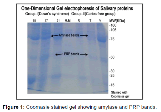
Protein separated out according to their relative mobility in gel and tinted patterns according to the criteria described by Azen et al. [14] The number of tinted bands present for each subject was counted on the CBB (Coomassie Brilliant blue stain); PRP-1 were marked according to band size and stain vigour as absent (–), present (+), and high intensity and size (-). However, due to subjectivity inherent in these parameters, salivary molecules were additionally scored only as present and absent as shown in Figure 1.
A specially designed format was used to record the personal data as well data related to measured variables. . The data so obtained was compiled, tabulated, described and statistically analyzed using chi square test in order to arrive at the conclusions as shown in Table 1.
| Group | PRP Bands | χ2 - value | P-value | ||
|---|---|---|---|---|---|
| Present | Absent | Total | |||
| n (%) | n (%) | n (%) | |||
|  I (Down's syndrome) | 13 (87) | 2 (13) | 15 (100) | 0.370 | 0.543 NS |
|  II (Caries free Children) | 14 (93) | 1 (7) | 15 (100) | ||
Table 1: The p-value obtained on comparing both the groups was 0.370 (= 0.05) suggesting that Down's syndrome group and caries free children have similar number of proline rich proteins.
Results
Inter-group comparison of proline rich proteins
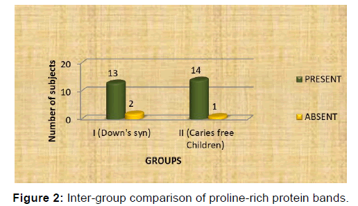
PRP bands were observed in 13 (87%) subjects in group I (Down’s syndrome) and in group II (caries free group) bands were observed in 14 (93%) subjects. The chi-square test was carried out. The p-value obtained on comparing both the groups was 0.370 (≥ 0.05) which indicated that this difference was statistically not significant suggesting that Down’s syndrome group and caries free children have similar number of proline rich proteins as shown in Table 1 and Figure 2.
Discussion
The prediction of the risk of caries has been of time an honoured interest and is crucial for advancement of new preventive layouts for caries. The prevalence of caries among 5 to 15 year old children was reported to be 56.2% according to an extensive, comprehensive national health survey conducted in 2004 stated by Bagramian RA [15] Purohit BM stated that the prevalence of caries was higher in children with special health care needs, than in healthy controls. [16] This was due to their potential motor, sensory and intellectual disabilities limiting their oral hygiene performance. [17] But the peculiar and interesting finding is that the prevalence of dental caries has been reported to be very low among children with Down’s syndrome compared to other special children or normal children.
It has been reported that the reason for this might be different environmental factors, congenital oligodontia, delayed eruption of teeth and different salivary composition. However E Davidovich stated that acidity in the mouth, salivary buffer adequacy, number of bacteria, are almost similar in patients with Down’s syndrome and normal children. [18]
Although Saliva is a supreme component for the local host defence opposing caries and gum defects hence lack of its secretion contributes to disease process stated by Shafer et al. [19] However unlike whole blood, saliva is easy to collect, less painful to the patient, and is less infectious for the health care provider. Although saliva has not been used till date as a sampling media, it does have a strong potential for use in the same type of tests that are done currently using blood. [20]
As already framed by Dodds et al. that the unstimulated character is the predominant condition in terms of the salivary gland functioning, thereby unstimulated whole saliva of all the subjects was flocked for analysis in this trial. [21]
Electrophoresis separation exposed a considerable variation in patterns of different individuals as shown by substantial differences in number, intensity, and size of observed bands. In this study, the presence of genetic polymorphism in salivary proteins of human whole saliva can thus be acknowledged. The number of PRP bands in either of the groups is comparable. The existence of proline rich proteins was observed in a quantum of 27 subjects (13 Down’s syndrome subjects and 14 caries free children).
Siqueira et al. stated that salivary protein concentration was 36% higher in Down’s syndrome patients than normal children. [22] Contradictory to such studies Sortino et al. observed no difference in protein concentrations between saliva samples of subjects with Down’s syndrome and control group. [23]
Similar to present study, Shobha tandon et al. observed proline rich proteins in caries free children in 53% subjects [24] and Banderas-Tarabay et al. observed PRP bands in 95% subjects. [25] Beeley suggested that this might be due to few pink staining PRP bands. It has been elucidated by the same author that the liability of the pink-violet staining PRP bands is a reflection of the unpredictable loss of these proteins. [26]
Bennick et al. [27] stated that Proline-rich proteins are chief constituents of parotid and submandibular glands saliva in humans as well as other animals. They can be further divided into acidic, basic and glycosylated proteins. The acidic proline-rich proteins will adhere to calcium to maintain the concentration of ionic calcium in saliva. Likewise they can inhibit crystallization of hydroxyapatite. Cowman RA observed limited growth of S. mutans in saliva from CARIES FREE individuals may be relatable to more than one factor. The protein composition of saliva may be a determinant of the oral microbial ecology of an individual and, by extension, of his inherent susceptibility or resistance to dental caries. [28] Basically, the vital PRP’s in the whole saliva residues neutralize acid from carbohydrate metabolism in situ (within the biofilm). The bulkier the concentration of available basic PRP’s, the greater will be no. of basic residues adherent to acid-producing streptococci and therefore the more efficient the neutralization. [29]
Conclusion
Contemporary dental caries research seeks to categorize risk factors as well as natural oral defenses that may protect against or prevent dental caries development. Saliva, in spite of being the strongest defense system, still has a wide array of properties and proteins whose role is yet not clearly known. Existing literature says that PRP’s might play a pivotal role in protection against dental caries which paves a way for the use of salivary proteins, like PRP’s as biomarkers in prediction of individuals at high risk for dental caries. On gel electrophoresis, in the present study there was a significant difference noticed among both groups with caries-free subjects having a higher number of proline-rich protein bands, substantiating the protective role of this protein. However further studies are warranted with greater number of subjects for more reliable and conclusive results. Further detection by monoclonal antibody or LC/MS analysis and other assays to support the result at nano level which escort to burnish the potentiality of its use in caries prediction is to be established through extensive research.
Conflict of Interest
All authors disclose that there was no conflict of interest.
REFERENCES
- Park K. Textbook of Preventive and Social Medicine, 19th ed, Banarsidas Bhanot publishers 2007: 467.
- Desai SS. Down syndrome: A review of the literature. Oral Surg Oral Med Oral Pathol Oral Radiol Endod 1997; 84: 279-85.
- Regezi J, Sciubba J. Oral pathology clinical pathologic correlations 1st edition In:Regezi, Sciubba editors. Philadelphia:WB Saunders Co 1989; 450-451.
- Gullikson JS. Oral findings in children with Down syndrome patients. Spec Care Dentist 1991; I: 248-251.
- Siqueira WL, Siqueira MF, Mustacchi Z, Oliveira ED, Nicolau J. Salivary parameters in infants aged 12 to 60 months with Down syndrome. Spec Care Dentist 2007; 27: 202-205.
- Allison PJ, Hennequin M, Faulks D. Dental care access among individuals with Down syndrome in France. Spec Care Dentist 2000; 20: 28-34.
- Oliveria AC, Paiva SM, Campos MR, Czeresnia D. Factors associated with malocclusions in children and adolescents with Down's syndrome. Is J Orthod Dentofacial Orthop 2008; 133: e1-e8.
- Asokan S, Muthu MS, Sivakumar N. Oral findings of Down's syndrome children in Chennai city, India. Indian J Dent Res 2008; 19: 230-235.
- Singh V, Arora R, Bhayya D, Singh D, Sarvaiya B, Mehta D. Comparison of relationship between salivary electrolyte levels and dental caries in children with Down syndrome. Journal of Natural Science, Biology, and Medicine. 2015; 6: 144.
- Helmerhorst EJ, Oppenheim FG. Saliva: A dynamic proteome. J. Dent. Res 2007; 86: 680-693.
- Yarat A, Akyuz S, Koc L, Erdem H, Emekli N. Salivary sialic acid, protein, salivary flow rate, pH, buffering capacity and caries indices in subjects with Down's syndrome. J Dent 1999; 27: 115-118.
- Thylstrup A, Fejerskov O. Textbook of Clinical Cariology, 2nd ed, Munksgaard 1994: 28-35.
- Schwatz SS, Zhu WX, Sreebny LM. Sodium dodecyl sulphate polyacrylamide gel electrophoresis of human whole saliva Archs Oral Biol 1995; 40: 949-958.
- Azen EA. Genetic protein polymorphisms in human saliva: An interpretative review. Biochem Genet 1978; 16: 79-82.
- Bagramian RA. Global increase in dental caries a pending public health crisis Am J Dent. 2009; 22: 3-8.
- Purohit BM, Singh A. Oral health status of 12 year old special children and healthy controls in southern India WHO South East Asia J Public Health 2012; 1: 330-338.
- Sanjay V, Shetty SM, Shetty RG. Dental health status among sensory impaired and blind institutionalized children aged 6-20 years J Into Oral Health 2014; 6: 55-58.
- Davidovich E, Aframian DJ, Shapira J, Peretz B. A comparison of the sialochemistry, oral pH, and oral health status of Down syndrome children to healthy children. International journal of paediatric dentistry. 2010; 20: 235-241.
- Shafer WG, Hine MK, Levy BM. 5th ed, Philadelphia: WB. Saunders Company A text book of oral pathology 1993; 567-658.
- John T, Devitt Mc. Saliva as the next best diagnostic tool. J Biochem. 2006; 45: 23-25.
- Dodds MW, Johnson DA, Mobley CC, Hattaway KM. Parotid saliva protein profiles in caries-free and caries-active adults. Oral Surg Oral Med Oral Pathol Oral Radiol Endod 1997; 83: 244-251.
- Siqueira WL, Nicolau J. Stimulated whole saliva components in children with Down syndrome Spec Care Dentist 2002; 22: 226-230.
- Sortino F, Pernicone C, Avitabile M, Vento M. Behavior of salivary proteins in patients with Down’s syndrome. Stornut of Mediterr 1985; 5: 81-84.
- Bhalla S, Tandon S, Satyamoorthy K. Salivary proteins and early childhood caries:A gel electrophoretic analysis Contemp clin dent 2010;1 (1):17-22.
- Banderas-Tarabay JA, Zacarías-D’Oleire IG, Garduño-Estrada R, Aceves-Luna E, González-Begné M. Electrophoretic analysis of whole saliva and prevalence of dental caries- A study in Mexican dental students. Arch Med Res 2002; 33: 499-505.
- Beeley JA. Clinical applications of electrophoresis of human salivary proteins. J Chromatogr 1991; 569: 261-280
- Cowman RA, Baron SS, Fitzgerald RJ, Danziger JL, Quintana JA. Growth inhibition of oral streptococci in saliva by anionic proteins from two caries-free individuals. Infection and immunity. 1982; 37: 513-518.
- Bennick. A Salivary proline-rich proteins Dept. of Biochemistry, University of Toronto, Toronto, M5S 1A8, Canada.
- Matsumoto-Nakano M, Tsuji M, Amano A, Ooshima T. Molecular interactions of alanine‐rich and proline-rich regions of cell surface protein antigen c in Streptococcus mutans. Molecular Oral Microbiology. 2008; 23: 265-270.


 The Annals of Medical and Health Sciences Research is a monthly multidisciplinary medical journal.